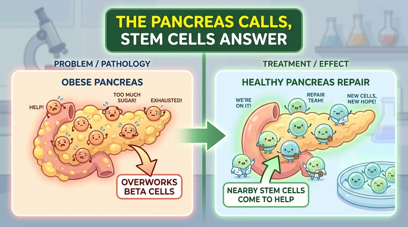
Why does being overweight lead to diabetes? Roughly speaking, it is because the body’s cells stop listening to insulin (insulin resistance). Even when the order goes out to lower blood sugar, the workers on the floor do not move. The troubled body tries to force the issue by shouting the order even louder. The factory that issues that order — that is, that puts out insulin — is the beta cell, found inside the small clusters of cells scattered through the pancreas: the pancreatic islets (islets of Langerhans).
For as long as obesity continues, beta cells work desperately. Not only does each one raise its own output; they also increase their sheer numbers to boost total production capacity. This gallant effort to hold the line is called beta cell compensation. As long as this compensation keeps up, blood sugar is somehow kept normal. Conversely, only when this effort reaches its limit and the factory finally gives out does type 2 diabetes (T2D) make its appearance. That is why beta cell compensation is the last seawall that decides whether or not you fall into diabetes.
And yet — exactly how is this seawall maintained? The molecular-level mechanism had long remained poorly understood. Are beta cells increasing their numbers all on their own? Or are they borrowing someone’s help? The paper introduced today gives a surprising answer to this question. A cornered beta cell releases tiny “parcels” and calls for help from the stem cells right next door — that is the story.
Journal information
- Paper title: Mesenchymal stem cells receive adaptive islet–derived miR-151–containing sEVs to promote β cell compensation in obesity
- Authors: Xinwei Guo (first author), Yang Wang, Ruixue Du, Wei Yong, Wenjing Yan, Zicheng Zhang, Yi Pan, Yanfeng Zhang, Yumeng Shen, Yue Yang, Fangfang Zhang, Jianxing Liu, Wei Tang (corresponding author), Yue Liu (corresponding author), Liang Jin (corresponding author)
- Affiliations: State Key Laboratory of Natural Medicines / Jiangsu Key Laboratory of Druggability of Biopharmaceuticals, School of Life Science and Technology, China Pharmaceutical University (Nanjing, China) / Department of Endocrinology, Geriatric Hospital of Nanjing Medical University / School of Pharmacy, Ningxia Medical University
- Journal: Science Advances (American Association for the Advancement of Science, AAAS), 2026, Vol. 12, No. 29, eadu4196
- DOI / link: 10.1126/sciadv.adu4196
- Impact Factor: about 13.9 (latest Journal Citation Reports, approximate; Q1 in the multidisciplinary sciences). Science Advances is a fully open-access multidisciplinary journal published by AAAS (the American Association for the Advancement of Science) and is a sister journal to Science
- Open access: Yes. This paper is CC BY 4.0 (free to reuse and redistribute as long as the source is credited). It is an online-only, fully open-access journal launched in 2015, electronic ISSN 2375-2548, indexed in DOAJ
- From submission to publication: submitted September 25, 2025 → accepted June 9, 2026 → published July 17
- Data availability: the microRNA sequencing data for the sEVs are publicly available in the GEO database (accession number GSE307474)
- Ethics review: animal experiments = Ethics Committee of China Pharmaceutical University (approval number 2024-05-015) / collection of human serum = Ethics Committee of Zhongda Hospital, Southeast University (2018ZDSYLL132-P01, in accordance with the Declaration of Helsinki, with written informed consent obtained from all participants)
- Conflicts of interest: the authors state “none declared”
About the corresponding authors: This paper has three corresponding authors (three names carry an asterisk, and each has a listed contact email). Starting with first author Xinwei Guo, the core experimental team belongs to the School of Life Science and Technology, China Pharmaceutical University (CPU), and the person who single-handedly leads the lab — from conceiving and supervising the study to providing resources, overseeing the project, and writing the manuscript — is Liang Jin (金亮).
Liang Jin is a professor and doctoral supervisor (博士生導師) at CPU’s School of Life Science and Technology, and has also been selected as a Jiangsu Distinguished Professor (江蘇省特聘教授). He earned his PhD at CPU in 2006, served as a postdoctoral fellow at the City of Hope National Medical Center in the United States, and returned to CPU around 2014 to take up a professorship. His specialties are “non-coding RNA and the regulation of blood-glucose homeostasis” and “stem cells and tissue regeneration” — making him a researcher whose main battlefield has been precisely islet beta cells and glucose metabolism. Indeed, he has built up a body of work on exactly the same “obesity, islet beta cells, non-coding RNA” theme that is continuous with this paper: a study showing that a circular RNA (circGlis3) protects islet beta cells from dysfunction and apoptosis under obesity (Nature Communications, 2023, corresponding author), work on the lncRNA βFaar (Nature Communications, 2021), and the obesity-induced miR-455 (Diabetes, 2022). Even his contact address, ljstemcell@cpu.edu.cn (which includes “stem cell”), symbolizes the axis of his research.
Co-corresponding author Wei Tang (drtangwei@njmu.edu.cn) is a clinical researcher based in the Department of Endocrinology of the Geriatric Hospital of Nanjing Medical University (Jiangsu Province Geriatric Hospital), working in that department’s “Islet Cell Senescence and Function Laboratory.” Through work such as a clinical study of 3,840 Chinese type 2 diabetes patients aged 60 and over, stratified by diabetes duration (Frontiers in Endocrinology, 2022, corresponding author), Wei Tang has pursued the age-related decline of islet function, insulin resistance, and diabetic complications from the perspective of a clinician who sees older patients every day. This is the role that connects the basic research (CPU) to the reality of the clinic.
The other corresponding author, Yue Liu (刘悦), is a researcher at the School of Pharmacy, Ningxia Medical University (in the pharmacology division). In fact she is part of Liang Jin’s lineage: she earned her PhD at CPU’s School of Life Science and Technology in 2023, and served as first author on the aforementioned circGlis3 paper (Nature Communications, 2023). Her current themes are drug discovery for the neurological complications of diabetes and neuroprotection against cerebral ischemia, and she has secured a National Natural Science Foundation grant (No. 82300906). In other words, someone trained in the stream of islet research from the Liang Jin lab has joined this collaboration from a different university.
In short, this paper is a collaboration that combines three standpoints — “basic science (CPU: stem cells and vesicle biology) × clinical medicine (Nanjing Medical University: geriatric endocrinology) × pharmacy (Ningxia Medical University)” — together with a web of people centered on the Liang Jin lab. (Titles and careers are given only to the extent that could be corroborated from public information; matters that could not be confirmed are not written on the basis of speculation.)
👦 Student: Dr. Exotaro, what exactly is an “exosome” or a “vesicle” to begin with?
🧬 Dr. Exotaro: It is a tiny “parcel” that a cell throws out into its surroundings, roughly 30–150 nanometers in diameter (one nanometer is one-millionth of a millimeter). Academically it is called a small extracellular vesicle (sEV). Inside it are packed proteins and RNA, and it acts like a courier delivering messages from cell to cell. The most important point today is this: “a parcel put out by a beta cell reaches another cell and ends up changing that cell’s character.”
What was it that we didn’t understand until now?
Who supports the beta cells’ “effort to hold the line”?
To repeat, when obesity makes insulin less effective, beta cells compensate by increasing their numbers and raising their secretion. During this compensation, what is happening inside the islet? Traditionally this tended to be described as a kind of self-sufficient picture, in which “beta cells cheer themselves on and multiply on their own.” But islets are not made of beta cells alone. They are a tiny society in which many supporting players coexist: endothelial cells that build blood vessels, macrophages that play the cleanup role, and mesenchymal stem cells (MSC), the backstage crew of the tissue, among others. Might these supporting players have a hand in compensation? That was a question that lay wide open.
The clue was the “vesicle”
What has become clear in recent years is that sEVs put out by islets appear to be sending messages to distant organs. In the prior work cited by the paper, islet-derived sEVs influenced the insulin sensitivity of the liver (miR-29), miR-155 in sEVs put out by macrophages inside the islet worsened glucose handling under a high-fat diet (miR-155), and islet-derived miR-204 mediated communication with skeletal muscle — sEVs were already known as “liaison officers” between organs.
On the other hand, there is also a basis on the side of the mesenchymal stem cells (MSC). MSCs exist in nearly all tissues throughout the body and are cells involved in tissue repair and the maintenance of homeostasis. Reports had been accumulating that administering MSCs to diabetic model animals improved blood sugar and advanced beta cell regeneration, and that MSC therapy improved metabolism in patients with type 2 diabetes. Here the authors set up a bold hypothesis that ties these two threads into one: the sEVs put out by islets (beta cells) speak directly to MSCs inside or near the islet, win the MSCs over to their side, and thereby push beta cell compensation forward.
👦 Student: If they are right next door, wouldn’t it be enough to just poke them directly rather than going to the trouble of sending a parcel?
🧬 Dr. Exotaro: A good question. But the parcel has its advantages. You can seal an “instruction sheet” called a miRNA (microRNA) inside it and deliver it, in bulk, to a chosen recipient. And if it rides the bloodstream, it can be carried far away. In other words, an sEV is a versatile means of communication that can serve both as an internal phone call to the folks next door and as a letter to somewhere distant. The crux of today’s story is that this parcel was being used addressed to “the MSCs in the neighborhood.”
The blank that had not been filled in
To sum up, the blanks at the starting point of this research were the following three. ① How do the sEVs of compensating beta cells under obesity differ from those under normal conditions? ② Which cells do those sEVs reach, and what do they make them do? ③ What is the true identity of the “cargo” doing the work, and which switch does it press inside the cell that receives it? This paper answers this three-tiered question, one step at a time.
What did this paper find?
① Beta cells during compensation were “overproducing” sEVs
First the team started by reproducing an islet in which compensation is occurring. When they extended the period for which wild-type mice were given a high-fat diet (HFD / 60% fat), impaired glucose tolerance, hyperinsulinemia, and hyperlipidemia progressed in stages, and by 9 weeks the islets had grown larger and beta cell division had become conspicuous. They treat this as the islet of the “compensation phase.”
They then compared sEVs harvested from normal islets (nid-sEVs: non-adaptive islet–derived) with sEVs harvested from compensating islets (aid-sEVs: adaptive islet–derived). The harvesting method here is as follows: the whole islets are isolated from the pancreas (they are not dispersed into single cells), cultured for about 48 hours in medium from which serum had been removed (sEV-depleted FBS), and the supernatant — that is, the components secreted into the culture medium — is collected. After removing cells and debris with staged centrifugation (300g → 2,000g → 10,000g), the sEVs are pelleted by two rounds of ultracentrifugation (120,000g), and in the later experiments they are further purified by size-exclusion chromatography (SEC) to remove contaminants such as free protein. In nanoparticle tracking analysis, both were in the typical sEV size range of 50–150 nm, and the compensating islets clearly released more sEVs. Size and shape (transmission electron microscopy) did not differ between the two; only the amount had increased.
That these vesicles are truly sEVs was confirmed by the detection of marker proteins (ALIX, TSG101, CD63, CD81) and by the absence of Grp94, an endoplasmic-reticulum protein that serves as a sign of intracellular contamination. In addition, beta cell–specific insulin and miR-375 were found inside the sEVs, and an experiment breaking the membrane with detergent showed that the insulin was enclosed inside the vesicles. This is a reminder that “this parcel does indeed come from beta cells.” RAB11A, one of the pieces of machinery that produce sEVs, was also increased in the compensating islets.
As the clincher, using a device that makes only beta cells glow (an AAV reporter expressing CD63-EGFP under the promoter of the insulin 2 gene), they tracked the fact that beta cell–derived sEVs are released into the blood (note, however, that the aid-sEVs collected in culture are themselves secretions of the whole islet, and this reporter and beta cell markers corroborate that a beta cell–derived fraction is included among them). Furthermore, in human serum as well, people with higher total cholesterol (>5.2 mM) or triglycerides (>1.7 mM) had more sEVs in the blood, and sEV amount showed a positive correlation with cholesterol and triglyceride values. This is supporting evidence from both the animal and human sides.
② That parcel was reaching the “islet MSCs”
The next question is which cells the increased sEVs reach. When compensating islets were dissociated and examined cell by cell, the fluorescent beta cell–derived sEVs had been taken up by all three types — CD73⁺CD90⁺ MSCs, CD31⁺ endothelial cells, and F4/80⁺ macrophages. At the whole-body level, labeled aid-sEVs mainly accumulate in the liver, lungs, and spleen.
What is interesting here is the difference in the mode of uptake. When they used a drug (cytochalasin D) that blocks “endocytosis” — the process by which a cell invaginates its membrane to swallow something whole — uptake decreased in endothelial cells and macrophages, but the effect was weak in MSCs. Conversely, when they applied a treatment that shaves off the surface proteins of the cell (proteinase K), only the uptake by MSCs weakened. In other words, macrophages and endothelial cells take up sEVs by “swallowing whole,” whereas MSCs receive them through a handshake between surface proteins — that is the difference.
So what is the molecule responsible for that handshake? The team comprehensively examined the proteins of the purified aid-sEVs by mass spectrometry (MS). Mass spectrometry is a technique in which the proteins contained in a vesicle are first chopped into short fragments, and the weight and sequence of each fragment are read precisely and matched against a database to work out “which protein it originally was.” The resulting “parts list” ran to 4,637 kinds, of which the membrane proteins that lie on the vesicle surface numbered 34. Since the handshake agent must be a molecule showing its face on the outside, they could first narrow things down to these 34. Among them, the only candidates reported to be involved in sEV transport were three: STXBP1, VAMP2, and F11R. When they harvested sEVs from beta cells with each of these knocked down and tested them, only the sEVs lacking F11R became harder for MSCs to take up. Conversely, knocking down F11R on the MSC side also drastically reduced uptake. F11R (F11 receptor, also known as JAM-A, a type of adhesion molecule) forms the bond by having the molecules on the vesicle side and the receiving side shake hands — that was the true identity of the selective handoff to MSCs.
👦 Student: If it reaches three types of cells, why is the MSC the lead role?
🧬 Dr. Exotaro: That is exactly the point of the next experiment. “Reaching” and “having an effect” are two different things. Even if you hand out letters to three people, only one might write back and take action, right? The team went precisely to pin down “which cell actually takes action.”
③ The one “producing the effect” was the MSC
So the team had MSCs, endothelial cells (MS1), and macrophages (RAW264.7) each take up aid-sEVs, and then co-cultured them with islets or beta cells (MIN6) in a vessel partitioned by a membrane (a Transwell). Because there is a partition, this is a setup in which they exchange only secretions without touching directly.
The result was clear-cut. The only ones that promoted beta cell division and raised glucose-stimulated insulin secretion (GSIS) were the MSCs that had taken up aid-sEVs. Having endothelial cells or macrophages take up aid-sEVs did not produce this effect. It was the same in whole mice: obese mice given MSCs loaded with aid-sEVs showed lower fasting blood glucose, increased insulin levels, advanced beta cell division, and a decrease in HOMA-IR, an index of insulin resistance. The role of pushing beta cell compensation forward was carried by the MSC.
What is more, aid-sEVs even changed the MSCs’ own character. MSCs bathed in aid-sEVs divided more actively (EdU assay), and the total amount of protein they secreted into the culture medium increased. When the secreted material was subjected to mass spectrometry, the pathway most strongly enriched was the WNT signaling pathway. In fact, the expression of several WNT genes went up, and among them the proteins WNT3 and WNT3A clearly increased by ELISA as well. WNT is a representative growth signal that promotes cell proliferation. In other words, the MSCs that received aid-sEVs secreted WNT3/WNT3A and sent the order “multiply” back to the beta cells — the players and the props were all in place.
④ The identity of the cargo — miR-151
So what is it in the aid-sEVs that changes MSCs this much? sEVs carry both proteins and RNA, but the team first checked whether “the miRNA is what’s working.” When they harvested sEVs from beta cells rendered unable to make miRNA (with Dicer knocked down), those sEVs lost their effect. Turned around, this means the lead actor doing the work is the miRNA.
So when they compared the miRNAs of aid-sEVs and nid-sEVs, 61 kinds were increased and 15 kinds were decreased. The most abundantly contained was miR-151. This miR-151 was also increased in the islets of high-fat-diet mice, and it likewise increased when cultured beta cells were stimulated with palmitic acid (a kind of fatty acid). Its position as “an instruction sheet that increases in the emergency of compensation” becomes clear.
The decisive part was the following separation. When they made an sEV artificially loaded with more miR-151 and an sEV with less and compared them, only the sEV with more raised beta cell division and insulin secretion, while the sEV with less had no effect. In whole mice as well, administering sEVs loaded with more miR-151 improved glucose tolerance and insulin sensitivity, and raised blood WNT3/WNT3A. Even more carefully, using size-exclusion chromatography (SEC) to separate vesicles from dissolved protein, they confirmed that miR-151 is concentrated on the vesicle side and that the effect too is on the vesicle side. “miR-151 is carried to MSCs riding the vehicle called an sEV” — they nailed it down this far.
There is also an interesting side note. Forcibly expressing miR-151 in the beta cells (MIN6) themselves did not change beta cell division or insulin secretion. miR-151 does not act directly on beta cells; it only works once it has first been handed over to the MSC. An instruction sheet sent to the wrong address does not work.
👦 Student: What can such a short RNA as miR-151 actually do?
🧬 Dr. Exotaro: A miRNA is a small RNA of just over 20 letters, but it has the job of taking aim at a specific gene and “silencing” it. It is just like a small sticky note that switches off only one particular switch on a machine. The next story is about which switch inside the MSC that note switched off.
⑤ The switch pressed inside the MSC — a brake called KLF9
The team narrowed down the partner that miR-151 targets inside the MSC using prediction tools (miRWalk and JASPAR), and finally arrived at a gene called KLF9 (Krüppel-like factor 9). KLF9 is a transcription factor — that is, a “switch operator” that decides whether other genes are on or off.
Note the direction here. KLF9 is a transcription factor that plays the brake role, suppressing MSC proliferation and WNT secretion. In fact, when KLF9 was forcibly increased, both MSC division and WNT secretion dropped. And in compensating islets, and in islets treated with aid-sEVs, the expression of KLF9 was lowered. Giving miR-151 to MSCs decreases KLF9. In other words, miR-151 was the sticky note that releases the brake called KLF9.
That this relationship is “a genuine direct action” was also carefully verified. At the end of the KLF9 gene (the 3′ untranslated region) there is a conserved sequence to which miR-151 binds (shared between human and mouse); a reporter incorporating that sequence had its activity lowered by miR-151, and when the sequence was destroyed it was no longer lowered. It was also shown that miR-151 and the KLF9 mRNA are captured together inside the machine that silences genes (the RISC, which contains Ago2). Conversely, knocking down KLF9 directly raised MSC proliferation and WNT secretion just as giving miR-151 did, and those MSCs promoted beta cell division and insulin secretion. Moreover, combining KLF9 knockdown with aid-sEVs does not add any extra effect — strong circumstantial evidence that the two lie on the same pathway.
Finally, what happens when the brake called KLF9 is released? KLF9 was binding to the promoters of two genes — Ccnd1 (cyclin D1), which drives proliferation, and Wnt3a, the growth signal — and suppressing them. So KLF9 coming off = the brakes on Ccnd1 and Wnt3a coming off. The MSC proliferates (Ccnd1) and secretes WNT (Wnt3a). Everything connected into a single line.
The whole picture — the “call for backup” that beta cells sent to stem cells
Drawing the whole series of results into a single picture, it goes like this.
- Beta cells cornered by obesity and insulin resistance overproduce and release sEVs loaded with miR-151.
- Those sEVs are handed selectively to nearby MSCs via the handshake molecule F11R.
- Inside the MSC, miR-151 silences KLF9 (the brake).
- With the brake released, Ccnd1 (proliferation) and Wnt3a (secretion) start moving, and the MSC proliferates and secretes WNT3/WNT3A.
- That WNT returns to the beta cells and pushes beta cell division and insulin secretion forward = compensation is supported.
Cornered beta cells were not going it alone; they were awakening the stem cells right next door as “helpers.” This circuit of “self-rescue” is the islet’s survival strategy that this paper has revealed.
How might the future change? (The road to the clinic)
Let us organize this discovery from a clinical perspective.
First, a new target for “protecting beta cells” has come into view. Conventional diabetes treatment centered on directions such as supplementing insulin, whipping beta cells to “put out more,” or easing peripheral insulin resistance. What this study showed is a path in which the body’s own seawall of beta cell compensation might be reinforced from the outside. And the target is not the beta cell itself, but its helper, the MSC. This connects to a shift in thinking: increasing the support corps is more sustainable and gentler than directly whipping the beta cells.
Second, three concrete molecular targets have come to hand. miR-151 (increase it), KLF9 (suppress it), and delivery to MSCs via F11R — manipulating any point in this circuit could, in principle, push beta cell compensation forward. In particular, the fact that the “cargo (miR-151)” and the “address (F11R)” have been identified connects directly to a concrete drug-design concept: an engineered exosome loaded with miR-151. It is a bridge from the stage of “using stem cells somewhat vaguely” to the stage of controlling “which molecule to deliver, to which cell, and how.”
Third, the urgency of the target. Type 2 diabetes keeps increasing worldwide, and at its heart lies the collapse of beta cell compensation. If we can provide support before compensation breaks, or just as it is beginning to break, the significance of delaying the onset and progression of diabetes is large. In this study, administering aid-sEVs or sEVs loaded with miR-151 improved not only beta cell mass but also whole-body metabolism in the form of glucose tolerance and insulin sensitivity.
On the other hand, the hurdles to reaching humans are high. In addition to the standardization of dose, route of administration, and manufacturing (how to guarantee the quality of miR-151 content and F11R), even in this study the aid-sEVs mainly accumulated in the liver, lungs, and spleen, and could not target the pancreas specifically. How to deliver them to the islets remains unsolved. Furthermore, aid-sEVs are taken up not only by MSCs but also by macrophages and endothelial cells, and the downstream effects there are not understood. And the greatest reservation is that this is a result at the preclinical stage using cells and mice, and clinical trials in humans have not yet been carried out.
How to read this study critically — the limitations, and the road to raising its quality further
The better a paper is, the more meaning there is in accurately grasping its limitations. First, let us fairly list the points where this study excels.
- Multilayered, thorough verification. Stacking together whole animals (mice), cultured cells, co-culture, proteomics, miRNA sequencing, and transcriptomics, they take down one by one — with both the wheels of genetics (knockdown, mutant reporters, Ago2 immunoprecipitation) and functional experiments — the delivery (F11R), the cargo (miR-151), the target (KLF9), and the downstream (Ccnd1/Wnt3a). Rather than stopping at “it worked,” the completeness with which they drew out the entire circuit to the end is high.
- Incorporating human specimens. Not with mice alone; they take supporting evidence for the sEVs in human serum (correlation with total cholesterol and triglycerides).
- Identifying both the “address” and the “cargo.” Simultaneously securing the two keys of delivery and effect — F11R and miR-151 — is of great value when thinking about therapeutic application.
On that basis, let us list the limitations.
① Verification mainly in male mice. The whole-animal experiments, including the compensation experiments, center on male C57BL/6J mice. Clear sex differences are known in obesity, glucose metabolism, and beta cell responses, and it is not clear whether the same circuit works in females. Verification in contexts where sex hormones greatly move metabolism, such as aging and pregnancy, is also a task for the future.
② “Beta cell compensation” could be a double-edged sword. This study portrays “supporting” compensation as good, but theoretically the intensification of compensation could also hasten the exhaustion and depletion of beta cells over the long term. Whether short-term improvement in blood sugar protects beta cells or wears them out in the long view cannot be concluded without long-term follow-up.
③ The safety of increasing miR-151. miR-151 is “a miRNA that increases with obesity,” and prior work has shown that its blood concentration is high in people with prediabetes and insulin resistance. In other words, it is a molecule that also has the aspect of a “marker of the disease state” to begin with. Moreover, because one miRNA targets many genes, increasing miR-151 throughout the body could also act on targets other than KLF9. Whether the direction of increasing it as a therapy is truly safe requires careful judgment.
④ The specificity of delivery is “partial.” The selective uptake into MSCs is explained by F11R, but as the authors themselves acknowledge, of the 34 membrane proteins identified, only some — including F11R — were verified, and the contribution of other receptors is a remaining question. The role of aid-sEVs in the macrophages and endothelial cells into which they are taken up (actions other than beta cell compensation) also remains unexplained.
⑤ The reality of the cell population called “islet MSCs.” The MSCs in this study are, so to speak, an operational population defined by surface markers (CD73⁺CD90⁺CD31⁻CD45⁻) from cells that crawled out of the islet. There is room to further pin down, through lineage tracing and the like, how many “islet-resident MSCs” exist in the living body and in what form, and whether their properties have changed in culture.
⑥ The transparency of statistics and design. The analyses are mainly t-tests and analysis of variance, and the sample sizes are roughly n=3–6 — standard for this kind of study, but not large-scale. Descriptions of preregistration and blinding of assessors are sparse, and there is room for improvement in terms of reporting transparency.
⑦ “Islet-derived sEVs” are, in reality, secretions of the whole islet. The aid-sEVs / nid-sEVs used in the functional experiments were collected from the supernatant of cultured whole islets, and are a mixture of sEVs from all cell types — not only beta cells, but also alpha cells, delta cells, endothelial cells, macrophages, and MSCs. The attribution “beta cell–derived” is indirect corroboration based on detection of a beta cell–specific reporter (Ins2-CD63-EGFP) and beta cell markers (insulin, miR-375), and does not mean that only beta cells were sorted out and collected. Therefore, whether miR-151 is truly released riding on beta cell sEVs, and how large the contribution of other islet cells is, is not strictly separated.
So then, how could this have become an even higher-quality study? Here are concrete proposals.
- Both sexes + larger group sizes, a design based on prior power calculation, and explicit blinding of assessors and adherence to ARRIVE.
- MSC-specific conditional knockout of KLF9 in the living body, to directly prove that the “miR-151 → KLF9 → WNT” axis is necessary for beta cell compensation (at present the evidence is mainly circumstantial — correlation plus the absence of an additive effect).
- Presentation of a delivery technology that targets the islets (targeted engineered exosomes) and a dose–response relationship. An answer is needed to the problem that systemic administration is biased toward the liver, lungs, and spleen.
- Long-term follow-up to determine whether pushing compensation forward protects beta cells or exhausts them.
- A safety assessment of the systemic action of miR-151 (effects on other organs and other targets).
- Identification of islet-resident MSCs in the living body (lineage tracing, spatial analysis).
- Direct isolation and analysis of beta cell–derived sEVs (such as sorting of reporter-labeled sEVs) to separate the fact that miR-151 is being put out riding on beta cell sEVs from the contribution of other islet cells.
Dr. Exotaro’s perspective
To be honest, diabetes and beta cell metabolism are not my own field of specialty. What I myself have long worked on is the theme of how to use mesenchymal stem cells (MSC) and their extracellular vesicles (EV) in the treatment of neurological diseases, starting with spinal cord injury (SCI). Both the organ and the disease state differ from this paper.
Even so, while reading it I found myself slapping my knee again and again. The reason is that this study turned the common sense of our own field on its head.
In our field, we think in terms of a scheme in which the EVs put out by MSCs are delivered into the body as a “drug” to help injured nerves. The MSC-EV is the sender, and the injured tissue is the receiver. But in this paper, that relationship was reversed. The sender of the EVs was the cornered beta cell, and the MSC was the receiver. And that MSC was not an administered drug, but a cell that resides in the body from the start. The body is mobilizing MSCs on its own, as an “amplifier,” in response to an SOS from a target organ — this idea was a fresh surprise to me. The MSC is at once a tool we administer from the outside, and also a responder cell that the body naturally relies on.
The second thing I learned is that you can rewrite the secretions of a helper cell (its secretome) with just a single miRNA. Simply by miR-151 releasing one brake called KLF9, the MSC switched into a “support mode” in which it proliferates and secretes WNT. In our field too, we keep contriving ways to load MSC-EVs with a specific miRNA and have them put out secretions favorable to neuroprotection and regeneration. That “reprogramming of the secretome” — nature was pulling it off with just a single sticky note. The four-tiered line of argument — effect → cargo → address → rewriting of the receiver — is the very path we should aim for in the neurological field.
Third is a shared carefulness. This study’s reservation that “compensation may be a double-edged sword” overlaps closely with the problem we face in the neurological field — that “good plasticity and an overshooting reaction are a hair’s breadth apart.” It is not a matter of just push and it’s good, just increase and it’s good. I was reminded anew that the control of when, where, and how much is precisely the heart of delivering regenerative medicine to the clinic.
Of course this is preclinical research, and efficacy in humans has not been shown. The safety of using miR-151 as a therapy is also a task for verification from here on. Even so, this study — which drew out, in the language of molecules, the circuit by which a cornered organ quietly calls for help from the stem cells next door — is one that, across organ boundaries, gives those of us involved in regenerative medicine much to reflect on.