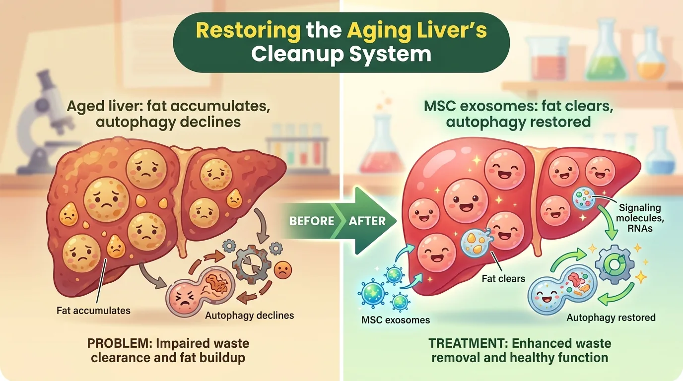
Plenty of people have been told at a routine check-up that they have a fatty liver. And the older you get, the more likely you are to hear it. You are not eating any more than you did in your twenties, yet somehow fat has accumulated in the liver — this is not simply a matter of self-indulgence. It is a biological phenomenon in which aging itself rewires the metabolism of the liver.
So what exactly breaks down inside an aging liver? Today’s paper points to autophagy, the process by which a cell wraps up its own waste, breaks it down and rebuilds from the parts — in effect, a recycling plant inside the cell. This plant rusts with age, and fat begins to outpace its capacity. As the “mechanic” that might restart the rusted plant, the research team turned to exosomes released by mesenchymal stem cells derived from the human umbilical cord.
Journal Information
- Article title: Mesenchymal Stem Cell-Derived Exosomes Improve Aging-Related Changes in Liver Lipid Metabolism by Enhancing Autophagy
- Authors: Jinquan Li (first author), Mengqi Gao, Jinke Feng, Dini Huo, Rui Hong, Fuhua Zhang, Qin He (corresponding author), Ming Dong (corresponding author)
- Affiliations: Department of Endocrinology and Metabolism, Qilu Hospital of Shandong University (Jinan, China), plus the Shandong Key Laboratory for Spatiotemporal Regulation and Precision Intervention of Endocrine and Metabolic Diseases, the Shandong Engineering Research Center for Advanced Technologies in the Prevention and Treatment of Chronic Metabolic Diseases, and the Institute of Endocrine and Metabolic Diseases of Shandong University
- Journal: Aging Cell (Wiley / The Anatomical Society), 2026, Vol. 25, e70642
- DOI / link: 10.1111/acel.70642
- Impact Factor: approximately 7.7 (2025 Journal Citation Reports, approximate; the 5-year IF is about 8.9). It is a fully open-access journal published by Wiley as the official journal of The Anatomical Society, and sits in Q1 of the geroscience field
- Open access: Yes (CC BY — free to reuse and redistribute with attribution)
- Submission to acceptance: received 17 March 2026 → revised 2 July → accepted 11 July
- Funding / conflicts of interest: National Natural Science Foundation of China (82300892), Natural Science Foundation of Shandong Province (ZR2025MS1417, ZR2022MH182), China International Medical Foundation (2024-N-05-05). Conflicts of interest are declared as “none”
- Ethics review: animal experiments = Animal Ethics Committee of Qilu Hospital of Shandong University (DWLL-2022-090, stated to comply with the ARRIVE guidelines) / human umbilical cord collection = the same hospital’s ethics committee (KYLL-202008(KS)-201, with donor consent obtained)
About the corresponding authors: The corresponding authors of this paper are Qin He (何芹) and Ming Dong (董明). Both are affiliated with the Department of Endocrinology and Metabolism at Qilu Hospital of Shandong University, and in addition hold positions at the three research platforms listed above (the Shandong Key Laboratory, the Shandong Engineering Research Center, and the Institute of Endocrine and Metabolic Diseases). Of the eight authors, these two are the only ones listed across all four institutions, which is consistent with a senior-author role. The author contribution statement likewise says that the two designed the study and reviewed and approved the manuscript, while first author Jinquan Li carried out the experiments and the writing.
Ming Dong (董明) is an MD, chief physician and doctoral supervisor (博士生導師), and deputy director of the hospital’s internal medicine teaching and research office. His specialty centres on pituitary and hypothalamic disorders, along with diabetes and its complications and thyroid disease. He spent a year as a visiting scholar at the Mayo Clinic in the United States, and serves on the Pituitary Group of the Endocrinology Branch of the Chinese Medical Association and on the Pituitary and Hypothalamic Disease Group of the China Alliance for Rare Diseases, among other roles.
For Qin He (何芹), what could be confirmed with certainty from public sources extends only to affiliation and research lineage; her clinical title could not be identified, so we refrain from writing anything speculative. The arc of the research, however, is clearly traceable. A 2018 first-author paper in Cancer Science on the AMPK-dependent mTOR pathway and autophagy (with Ming Dong as senior author) already shows the same “autophagy” axis that runs through the present paper. In a 2025 paper in Molecular and Cellular Biochemistry, He and Dong served in the corresponding-author role with the same Jinquan Li as first author, addressing NAFLD — evidence of a consistent interest in hepatic metabolism. She has also been a co-author on work linking MSC-derived exosomes to autophagy (AMPK/ULK1-dependent autophagy in diabetic muscle atrophy, Journal of Cachexia, Sarcopenia and Muscle 2023, among others), which places this group as one that has been building up the theme of “MSC exosomes × autophagy × metabolism”. It should be noted that two papers on MSC-derived exosomes on which He was first author and Dong a co-author (Stem Cell Research & Therapy, 2020 and 2021) were retracted in October 2022 because of problems with the figure images (both papers had a different researcher as corresponding author, and the retraction notices do not establish misconduct on the part of any individual author).
The Department of Endocrinology and Metabolism at Qilu Hospital, their home institution, was founded in the 1950s as the oldest specialist endocrinology and metabolism department in Shandong Province, and was designated a National Key Clinical Specialty in 2011 (77 staff, with over 94% holding doctoral degrees). Read in that context — a specialist department that sees, day in and day out, patients in whom fatty liver and impaired glucose metabolism accumulate with age, reaching for a tool from regenerative medicine as a way forward — the aim of this study comes into focus.
👦 Student: Exotaro, remind me — what is an “exosome” in the first place?
🧬 Exotaro: It’s a tiny “parcel” that cells release into their surroundings, roughly 30–150 nanometres across (a nanometre is a millionth of a millimetre). Inside are proteins and RNA, and it acts rather like a courier service delivering messages from cell to cell. The most important point for today’s story is that how well such a parcel works is decided by what cargo it happens to be carrying.
What Was Not Understood Until Now?
An aging liver quietly stores fat
The liver processes what we eat, stores sugar, remodels lipids and breaks down toxins — it is the hub of the body’s metabolism. Yet with age, fat becomes easier for this organ to accumulate. Medically this is called nonalcoholic fatty liver disease (NAFLD), and as it progresses it shifts into nonalcoholic steatohepatitis (NASH), which involves inflammation and cell injury, and from there towards fibrosis, cirrhosis and liver cancer. To make matters worse, there is still no established drug for NAFLD. The opening of the paper likewise notes that the mainstays of treatment are diet and lifestyle modification, and raises the absence of approved drugs as a “pressing challenge”. That is precisely why research hunting for new therapeutic targets is worth doing.
”Residents who won’t leave” — the phenomenon of cellular senescence
Another important thing happening in aged tissue is cellular senescence. It refers to a state in which a cell has stopped dividing and “retired” — but the trouble is that the retired cell does not leave the tissue; it stays put. And once settled in, senescent cells take on what is called the SASP (senescence-associated secretory phenotype), scattering inflammatory substances (TNFα, IL-6 and others) around them. The paper cites earlier work showing that the more senescent cells accumulate in adipose tissue, the higher the expression of the senescence markers P16 and P21, worsening liver injury through the release of free fatty acids and oxidative stress. In other words, cellular senescence and fatty liver make each other worse.
👦 Student: Retired but refusing to leave — that sounds like a nuisance.
🧬 Exotaro: Exactly. And if they just sat there quietly it wouldn’t be so bad, but they keep complaining to everyone around them. That’s the SASP. Which is why “can we reduce senescent cells?” has long been such a large theme in aging research.
The rusted “recycling plant” — autophagy
Now we come to autophagy itself. A cell wraps damaged proteins, worn-out mitochondria and surplus lipid droplets in a membrane to form a sac called an autophagosome, then fuses it with the lysosome (the organelle packed with degradative enzymes) to break the whole lot down. The resulting materials are reused — literally a recycling plant inside the cell.
In the liver, autophagy is also a key mechanism for breaking down lipid droplets (lipophagy). But according to earlier work cited in the paper, the efficiency of autophagy declines with age. When the production line slows, fat that cannot be processed piles up. The hypothesis that this is one cause of NAFLD is where this study begins.
Two molecules for measuring autophagy are worth memorising here, because they are key to the discussion that follows.
- LC3: a protein embedded in the autophagosome membrane. Its form before attaching to the membrane is LC3-I, and its form after attaching is LC3-II. Therefore a high LC3-II/I ratio = many autophagosomes are being made.
- P62: the “tag” attached to waste destined for degradation. If autophagy is turning over smoothly, it is degraded along with the tag and its level falls. In other words, rising P62 = degradation is stalled.
👦 Student: Wait — if P62 goes up, doesn’t that mean the cleaning is going well?
🧬 Exotaro: That is where the answer runs counter to intuition, and that’s what makes it interesting. Think of P62 as the rubbish bag itself. If the collection service is coming as it should, bags don’t pile up by the front door. If they are stacked in a heap, that’s proof the collection isn’t coming. So the accumulation of P62 is a sign that autophagy has stopped.
The gap that had not been filled
It had already been reported that exosomes derived from mesenchymal stem cells (MSC) ease liver fibrosis and drug-induced liver injury. But the authors point to the following gap. In a naturally aged state — not a model fattened on a high-fat diet, but an animal made old by the passage of time itself — can MSC-derived exosomes rebuild hepatic lipid metabolism? And by what mechanism? That question had been left almost entirely untouched.
What Did This Paper Find?
The tools used: human umbilical cord-derived MSCs and the exosomes harvested from them
The first thing the team prepared was human umbilical cord mesenchymal stem cells (HucMSC). Umbilical cords were donated from five full-term infants born by caesarean section to healthy young mothers, and cells were isolated and cultured from the “Wharton’s jelly” inside them. That the raw material is tissue that would otherwise be discarded as medical waste is a major advantage when thinking about practical application.
That the cells obtained were indeed MSCs was confirmed in two ways: by surface markers (over 99% positive for CD73 and CD105, and under 1% for HLA-DR and CD34 — matching the standard immunological definition of an MSC), and by their capacity to differentiate into both bone and fat.
The vesicles collected from the culture medium of these HucMSCs are the protagonists of this paper, HucMDEs (HucMSC-derived exosomes). Under transmission electron microscopy they were observed as spherical structures with a double membrane, and nanoparticle tracking analysis (NTA) gave a mean particle size of 144 nm — a typical size range for exosomes. On Western blot, the marker proteins HSP70 and TSG101 were more strongly enriched in the vesicles than in the parent cells.
Whether they really reach the liver was also checked. When HucMDEs labelled with Cy7 were injected via the tail vein, in vivo imaging showed them accumulating mainly in the liver. In culture dishes too, HucMDEs made to glow green with PKH67 were captured being taken up into the cytoplasm of the mouse hepatocyte line AML12.
① In naturally aged mice, metabolism and liver function improved
The experimental design is simple and solid. Male C57BL/6 mice were divided into three groups: a young group (Young, 8 weeks), an old group (Old, 18 months), and an old + exosome group (Old+HucMDEs). The key point is that the aging was natural, not driven by a high-fat diet. Dosing began at 18 months of age: HucMDEs at 5 µg per g of body weight, every three days for 12 weeks, injected via the tail vein. The old group received the same volume of PBS, and evaluation was carried out at 21 months of age (n=6 per group). The results were consistent.
- Body weight: the old group was heavier than the young group (p < 0.001), and HucMDEs significantly reduced it (p < 0.01)
- Intraperitoneal glucose tolerance test (IPGTT): the old group had a higher glucose peak that was slow to fall, with a large area under the curve (AUC) (p < 0.001). The HucMDEs group had a lower peak, returned nearly to baseline by 120 minutes, and its AUC was significantly reduced (p < 0.001)
- Intraperitoneal insulin tolerance test (IPITT): in the old group, glucose fell poorly even after insulin — that is, insulin resistance. In the HucMDEs group the fall in glucose was faster (nadir at 90 minutes) and the AUC was significantly lower (p < 0.01)
- Liver function: ALT and AST, indicators of liver injury, were elevated in the old group, whereas in the HucMDEs group they were significantly lower (p < 0.001)
- Blood lipids: both triglycerides (TG) (p < 0.01) and total cholesterol (TC) (p < 0.001) fell
The main indicators — body weight, glucose metabolism, liver function and lipids — all moved back towards the young end.
② Examining the liver itself showed less fat and less senescence
Blood tests alone cannot tell you what is going on inside. So the liver itself was examined histologically.
- H&E staining: liver architecture was disordered in the old group, whereas in the HucMDEs group the arrangement and morphology of hepatocytes partially recovered
- Oil Red O staining (which stains fat): the fat deposition that was heavy in the old group was reduced in the treated group
- SA-β-Gal staining (which stains senescent cells): the deep blue staining seen in the old group became fainter in the treated group
- PAS staining (which stains glycogen): hepatic glycogen, which had declined with age, increased in the treated group
Western blotting was then used to measure the amounts of the key proteins. This is a place where the direction is easy to get backwards, so let us lay it out carefully.
| Protein | Role | In the old group (Old) | With HucMDEs |
|---|---|---|---|
| SREBP1 | drives the synthesis of fat | up (p < 0.01) | down (p < 0.05) |
| PPARα | drives the breakdown (catabolism) of fat | down (p < 0.01) | up (p < 0.05) |
| P16 / P21 | markers of cellular senescence | up (p < 0.01) | down (p < 0.05) |
Remember that SREBP1 and PPARα always move in opposite directions. In an aged liver, the machinery that makes fat (SREBP1) grows stronger while the machinery that burns it (PPARα) weakens. HucMDEs pushed both back towards the young end.
③ The same picture in culture — with a control that did not work
The team also tested their in vivo results in cultured cells. The model adds palmitic acid (PA) to the mouse hepatocyte line AML12 to induce fat accumulation and cellular senescence (the concentration as written in the paper is 0.5 µM; we return to this notation in the critical reading below). What should not be missed is that they included a control exosome. Exosomes derived from human embryonic lung fibroblasts (HELF), called HEDEs, were prepared to separate “do exosomes of any kind work?” from “does it work because it comes from HucMSCs?”
The results were clear-cut. Against the fat deposition induced by PA, HEDEs did almost nothing, and only HucMDEs reduced it. The same held for senescence genes: P16, P21 and P53 rose with PA treatment but fell significantly when HucMDEs were added (p < 0.05). In the HEDEs group, by contrast, none of the three differed meaningfully from the PA group, and P21 and P53 were in fact slightly higher. The effect is specific to the HucMSC origin, as the data showed.
The behaviour of genes (mRNA) involved in lipid metabolism was also consistent with the in vivo results. Note that the following are mouse gene names, written differently from the protein names in the table above.
- Lipogenesis (Srebp1, Fasn): up with PA → down with HucMDEs (p < 0.001)
- Fat breakdown (Ppara, Cpt1a): down with PA → up with HucMDEs (p < 0.05)
- Triglyceride synthesis (Gpat1, Dgat2): up with PA (p < 0.05) → down with HucMDEs (p < 0.05)
- Lipid transport (Mttp, Apob): up in the HucMDEs group (p < 0.05)
“Suppress synthesis, promote burning, reduce triglyceride assembly, and increase export” — several pathways moved at once, all in the direction of not hoarding fat.
④ The heart of the matter: the rusted autophagy started turning again
From here on we are at the centre of the mechanism. Recall LC3 and P62 from earlier.
In vivo (mouse liver) — by both immunohistochemistry and Western blot, the old group showed increased P62 (p < 0.001) and a reduced LC3-II/I ratio. These are signs of stalled autophagy. In the HucMDEs group, P62 decreased (p < 0.05) and the LC3-II/I ratio rose significantly (p < 0.001). The paper honestly notes, however, that the levels did not fully return to those of the young group.
In cultured cells the picture was the same. PA treatment increased P62 (p < 0.001) and lowered the LC3-II/I ratio (p < 0.05). Adding HucMDEs decreased P62 (p < 0.001) and raised the LC3-II/I ratio (p < 0.001).
They also used RFP-GFP-LC3, a tool that distinguishes the “flow” of autophagy by colour. Green (GFP) is quenched in the acidic environment of the lysosome, so red alone means fusion has occurred (the flow is running), while green and red overlapping into yellow means fusion has not yet happened and things are stalled. The number of autophagosomes per cell was lower in the PA group than in controls, and significantly higher in the HucMDEs group than in the PA group (p < 0.01).
⑤ Testing whether autophagy is really necessary — by breaking it
Correlation alone cannot show that autophagy is the cause of the effect. So the team used four ways of breaking autophagy.
- siATG5 (knocking down the autophagy-related gene ATG5 with siRNA)
- siATG7 (knocking down ATG7 in the same way)
- 3-MA (3-methyladenine, 10 mM; inhibits the initiation of autophagy)
- Bafilomycin A1 (Baf, 30 nM; inhibits lysosomal acidification, i.e. stops the final step of degradation)
Under every one of these conditions, HucMDEs significantly raised the LC3-II/I ratio (p < 0.05 under 3-MA, p < 0.05 under Baf) and eased the accumulation of P62. In ATG5-knockdown cells, HucMDEs treatment partially restored ATG5 expression, though not to control levels (p < 0.01), while the LC3-II/I ratio was in fact pushed above the control level (p < 0.05). In ATG7-knockdown cells, both ATG7 expression and the LC3-II/I ratio recovered past the control level (both p < 0.001). The authors interpreted this as showing that “HucMDEs can rescue impaired autophagy whether it arises from gene knockdown, inhibition of initiation, or lysosomal dysfunction.”
⑥ Identifying the cargo — THBS1
Exosomes carry hundreds of different proteins. So which one is doing the work?
The team compared the proteins of HucMDEs and HEDEs comprehensively (proteomic analysis with DIA-NN). The two showed clearly different profiles, and among them one of the proteins most strongly enriched in HucMDEs was thrombospondin-1 (THBS1). In terms of the functions of the enriched protein set, “extracellular matrix (ECM) organisation”, “cell-matrix adhesion”, “lysosome”, “ECM-receptor interaction” and the “PI3K-Akt signalling pathway” ranked at the top. That lysosome-related pathways surfaced connects naturally to the autophagy story.
Then came the decisive experiment. THBS1 was knocked down in the HucMSCs with shRNA, and exosomes were harvested from those cells (the THBS1-knockdown version of HucMDEs) and put through the same tests. The result: exosomes lacking THBS1 could no longer reduce fat deposition, restore glycogen, or ease cellular senescence. Removing a single item of cargo abolished the effect — a persuasive result showing that THBS1 is the central effector molecule.
👦 Student: So taking one thing out of the parcel made it stop working?
🧬 Exotaro: Precisely. This is where the paper puts in its greatest effort. Rather than stopping at “the exosomes worked”, it went on to “this is the cargo that made them work”. If you want to use this as a therapy, that step is decisively important. Once you know the contents, you can also design an “enhanced” exosome loaded with more of that cargo.
⑦ The downstream switch — PPAR signalling
Finally, what happens where THBS1 arrives. The team performed RNA-seq on PA-treated AML12 cells with and without HucMDEs. 695 genes were differentially expressed (210 up, 485 down), and one of the most strongly enriched pathways in KEGG pathway analysis was the PPAR signalling pathway. In GO analysis, “extracellular exosome”, “cell surface” and “lysosomal membrane” also ranked at the top.
So they used GW6471, a drug that selectively inhibits PPARα. GW6471, like THBS1 knockdown, markedly attenuated the protective effect of HucMDEs. Fat deposition increased, glycogen stores fell, and senescence markers rose. PPARα is a downstream pathway required for HucMDEs to work — that is the conclusion.
That said, as the authors themselves carefully write, how THBS1 and PPARα are connected is still at the hypothesis stage. The paper sets out two possibilities: a route in which THBS1 initiates signalling via the receptor CD36 and converges on PPAR activation, or one in which the two act as separate parallel pathways that together create a metabolic environment favourable to autophagy. Which it is has been left to future experiments.
How Will the Future Change? (The Road to the Clinic)
Let us organise what this study shows from a clinical perspective.
First, it lends momentum to the strategy of “using the parcel rather than the cell”. Infusing stem cells themselves carries the risk of vascular obstruction, the problem of not reaching the intended site, immune reactions and concerns about tumour formation. Because exosomes are not cells, these can be structurally reduced. Moreover, the source material — Wharton’s jelly — is tissue that would otherwise be discarded after delivery, so it imposes no new burden on the donor.
Second, and most importantly, there is the single point that “the cargo has been identified”. Once you know that THBS1 is what works, the next design becomes concrete. Select exosomes loaded with plenty of THBS1, or load it in artificially. In the longer term, small-molecule drugs targeting THBS1 or the PPARα pathway are also conceivable. From the stage of “vaguely using stem cell culture supernatant” to the stage of controlling “which molecule moves which pathway, and how” — this study helps push that transition along.
Third, there is the urgency of NAFLD/NASH as a target. This field is short of established therapeutic drugs, and in an aging society patient numbers will only grow. What is more, this study improved more than just liver fat. Body weight, glucose tolerance and insulin sensitivity — whole-body metabolism — moved at the same time. The possibility of taking on age-related metabolic derangement as a package is clinically attractive.
At the same time, the hurdles before this reaches humans are high. Dose and dosing interval, route of administration, standardisation of manufacturing and quality control (how to guarantee particle size distribution, markers and THBS1 content), and long-term safety — THBS1 in particular is a pleiotropic molecule involved in the suppression of angiogenesis and in fibrosis, so the effects of long-term systemic administration must be assessed with care. Above all, no clinical trial in humans has yet been conducted. This study is a preclinical result obtained in cells and mice.
How to Read This Study Critically — Limitations, and Routes to Higher Quality
The better a paper is, the more it is worth understanding its limitations precisely. Let us first give fair credit to what this study does well.
- It used a natural aging model. In a field dominated by models that create fatty liver artificially with a high-fat diet, verifying the finding in animals actually aged to 18 months is significant.
- It included a control exosome that does not work (HEDEs). This is a high-quality specificity control that closes off the interpretation “any exosome will do”.
- It verified across multiple layers — in vivo, in cultured cells, in the proteome and in the transcriptome — and confirmed the downstream pathway twice over, with a genetic approach (THBS1 knockdown) and a pharmacological one (GW6471).
With that said, here are the limitations.
① Sample size, and a male-only design. n=6 per group is standard for this kind of animal experiment, but it is not large. More serious is that only male mice were used. Clear sex differences are known in lipid metabolism, fatty liver and cellular senescence, and whether the same results would appear in females is unknown. Generalising this as aging research requires verification in both sexes.
② The notation of the palmitic acid concentration. The methods section states “palmitic acid (PA; 0.5 µM)”. But the range normally used in experiments that induce lipotoxicity in hepatocytes is 0.1–0.5 mM (millimolar), so as written this is about one-thousandth of that. It is most likely a typographical error in the units, but this matters to readers attempting replication. Since the experiment itself does reproduce fat accumulation, there is no reason to doubt the results, but in terms of reporting accuracy it should be corrected.
③ The THBS1 → PPARα connection is a proposal, not a proof. This is a limitation the authors themselves state explicitly. The paper says that “the direct molecular interaction between THBS1 and PPARα requires further experimental verification”, and sets out both possibilities — convergence via CD36 and parallel pathways. Care is needed not to read this as proof of causation.
④ Interpretation of autophagic flux. In experiments using a lysosomal inhibitor such as bafilomycin A1, flux is properly assessed by comparing, as a pair, how much difference in LC3-II arises with and without the inhibitor. Simply lining up LC3-II/I ratios side by side across conditions cannot strictly distinguish “more is being made” from “less is being destroyed”. Adding flux observation with RFP-GFP-LC3 was an appropriate reinforcement, but the interpretation of the inhibitor experiments should be read with caution.
⑤ Sparse reporting of blinding. There is a statement that grouping was done “randomly”, but whether the quantification of tissue staining and Western blots was performed with assessor blinding is not made explicit. Judging staining intensity is prone to subjectivity, and the presence or absence of blinding bears directly on the robustness of the results.
⑥ “Which cells did they reach?” remains unresolved. This too is a limitation the authors raise themselves. The liver consists not only of hepatocytes but also Kupffer cells (resident macrophages), stellate cells and sinusoidal endothelial cells, and which cell type took up the HucMDEs and produced the effect has not been identified.
⑦ The distance from mouse to human. A 21-month-old mouse is not the same thing as an elderly human. The gaps in dose conversion, duration of administration, and the comorbidities and polypharmacy of real clinical practice are large, and it bears repeating that preclinical results cannot simply be extrapolated to humans.
So how could the study have been made even better? Here are concrete suggestions.
- Both sexes plus larger group sizes, with sample size set on the basis of an a priori power calculation. Aging × sex is one of the factors that most affects reproducibility in this field.
- Assessor blinding plus a pre-registered protocol. Registering the analysis plan in advance and disclosing ARRIVE compliance item by item would greatly increase the transparency of reporting.
- Quantify autophagic flux by paired comparison with and without inhibitor. Running bafilomycin-treated and untreated arms side by side for each condition and calculating the difference as flux would remove the ambiguity of interpretation.
- Verify the THBS1–PPARα connection directly. As the authors themselves note, co-immunoprecipitation (Co-IP) and luciferase reporter assays are needed. Adding knockout or inhibition of CD36 would allow the proposed pathway to be tested directly.
- Show a dose-response relationship. Comparing several doses rather than a single one would demonstrate dose dependence of the effect and help bridge to dose design in humans.
- Identify the cell type (co-staining fluorescently labelled exosomes with cell-type markers, or single-cell RNA-seq), and test THBS1-rich exosomes directly. Knockdown showed that removing it abolishes the effect. Showing the converse — that exosomes loaded with more THBS1 work more strongly — would make the causal case stronger still.
Exotaro’s Perspective
To be honest, hepatic lipid metabolism is not my own field. What I have worked on for a long time is how to use mesenchymal stem cells (MSC) and their extracellular vesicles (EV) to treat neurological disease, above all spinal cord injury. The organ and the pathology are both different from this paper’s.
Even so, I found myself nodding again and again, because the “pattern of thinking” used here is exactly the same as ours.
First, the commonality of the platform. Exosomes harvested from human umbilical cord-derived MSCs are, as a toolkit, essentially no different from what we have used for neural injury. The same tool drives lipid metabolism in the liver and post-injury repair in the nervous system. Put the other way round, this reinforces the understanding that MSC-EVs are not organ-specific drugs but a general-purpose platform that raises the baseline of how cells behave.
Second, autophagy as a point of contact. After injury to the central nervous system too, autophagy handles the disposal of damaged mitochondria and abnormal proteins, and determines whether neurons survive. The phenomenon of this housekeeping function declining with age or injury is shared across organs, and if EVs can push it back, the scope of application does not stop at the liver.
Third, and what I felt was most worth learning, is the willingness to go all the way to identifying the cargo. The weakness of MSC-EV research has long been that “we can see it worked, but we don’t know what worked”. Without knowing the contents you cannot draw up quality specifications, you cannot explain yourself to regulators, and you cannot guarantee reproducibility. This study narrowed down candidates by proteomics, showed necessity by knockdown, and went on to confirm the downstream pathway pharmacologically. This three-step structure of “effect → cargo → pathway” is exactly the route we should be aiming for in the neurological field.
Of course this is preclinical research, and efficacy in humans has not been shown. The link between THBS1 and PPARα has not moved beyond hypothesis either. Even so, the idea that we might be able to restore an aged organ’s “power to clean house” from the outside feels like great hope when facing the decline of organ function that comes with age.
We cannot stop growing old. But the flows that have stalled inside aged cells might just be set moving again — today’s paper was one that left me thinking so.