A very interesting paper has been published in Nature, so I would like to introduce it.

A somato-cognitive action network alternates with effector regions in motor cortex - Nature
A brief summary..
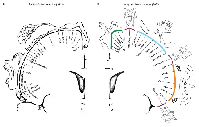
Do you know the brain map?
It is something like this.
This paper reports that reality is not as simple as this map; there are parts that coordinate with various other important networks.
This paper challenges the conventional view that the motor cortex is a continuous somatotopic homunculus, suggesting that the motor cortex is punctuated by the somato-cognitive action network (SCAN), a system for whole-body action planning. It was suggested that the inter-effector regions show decreased cortical thickness and are functionally connected to each other, as well as to the cingulo-opercular network (CON), which is essential for action and physiological control.
Explanation of terms
What is the somato-cognitive action network (SCAN)?
The Somatic Cognitive Affective Network (SCAN) is one of the brain’s functional networks and is involved in somatic, cognitive, and emotional processing. This network is composed of brain regions such as the prefrontal cortex, cingulate cortex, anterior temporal cortex, mid-frontal cortex, dorsolateral prefrontal cortex, and temporal pole.
The Somatic Cognitive Affective Network (SCAN) is one of the brain’s functional networks and is involved in somatic, cognitive, and emotional processing. This network is composed of brain regions such as the prefrontal cortex, cingulate cortex, anterior temporal cortex, mid-frontal cortex, dorsolateral prefrontal cortex, and temporal pole.
The somato-cognitive action network plays a role in linking emotional and bodily sensory information with cognitive processing, supporting higher-order functions such as self-awareness, understanding of others, and social cognition. This network is also involved in stress responses, self-regulation, and decision-making, and plays an important role in mental health and adaptation.
What is the cingulo-opercular network (CON)?
The Cingulo-Opercular Network (CON) is one of the brain’s functional networks and is involved in cognitive control and attention regulation. This network is composed of brain regions such as the anterior cingulate gyrus, posterior cingulate gyrus, prefrontal cortex, anterior insular cortex, and frontal eye field.
The cingulo-opercular network plays an important role in maintaining attention during cognitive tasks, adapting to tasks, detecting and correcting errors, and the decision-making process. This network also has the function of drawing attention to stimuli with high salience, and can selectively process information from the external environment and internal states.
Recent neuroscience research has revealed that the function of the cingulo-opercular network is associated with psychiatric disorders such as attention-deficit hyperactivity disorder (ADHD), schizophrenia, and depression. Research on this network is expected to contribute to understanding these disorders and developing treatments for them.
What is functional MRI?
Functional magnetic resonance imaging (fMRI) is one of the medical imaging diagnostic techniques for non-invasively observing brain activity. This technique uses a special magnetic resonance imaging device to indirectly observe brain activity by measuring changes in the oxygen concentration in the blood.
When the brain is active, blood gathers in that area. At this time, the oxygen concentration also changes, so fMRI can estimate brain activity by observing changes in the oxygen concentration in the brain. fMRI is used for the diagnosis and treatment of many disorders, including psychiatric and neurological diseases.
By having subjects perform specific tasks, fMRI can observe the activity of specific regions of the brain. For example, when a subject solves a math problem, activity in regions related to mathematics is observed. Also, when a subject is given a visual stimulus, activity in regions that process visual information is observed.
Because fMRI can observe brain activity non-invasively, it is widely used in fields such as neuroscience and neurosurgery.
How was the experiment conducted?
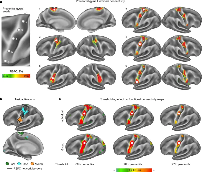
The methods used in this paper include precision functional magnetic resonance imaging (fMRI) to examine human motor cortex tissue. This study also used structural connectivity studies, non-human primate stimulation studies, and a series of motor and behavioral fMRI tasks to verify the findings. Furthermore, this study used data from the three largest fMRI datasets, as well as precision fMRI datasets of macaques and children (newborns, infants, and children), to analyze cross-species homologues and developmental precursors of the inter-effector system.
What results were obtained?
The results of this paper suggest that the conventional view of the motor cortex as a continuous somatic homunculus is incomplete. This study found that the motor cortex is intermittently formed with inter-effector regions that show decreased cortical thickness and strong functional connectivity to each other, as well as to the cingulo-opercular network (CON). These inter-effector regions are suggested to be part of the somato-cognitive action network (SCAN), a whole-body action planning system. This study also confirmed the existence of a concentric effector somatography demarcated by effector-specific (foot, hand, mouth) regions and CON-connected inter-effector regions.
What is effector somatography?
Effector somatography is a technique that records the firing patterns of motor neurons and visualizes the activity of muscles and other effectors. This technique is used as an important means of understanding the activity of the brain and nervous system.
Effector somatography can observe muscle contraction patterns by recording the electrical activity of motor neurons. This technique is used to examine muscle contraction patterns, the characteristics of electromyograms, and pathological abnormalities of motor neurons.
Effector somatography is widely used in fields such as the diagnosis and treatment of brain and nervous system diseases, motor learning, and sports science. It is also applied to the control of artificial effectors, such as the development of prosthetic limbs and robots.
What are the limitations of this study?
This paper has several limitations, including the fact that this study used only the fMRI method and did not investigate the motor cortex using other techniques such as direct cortical stimulation. Furthermore, this study did not investigate the functional significance of the inter-effector regions and their connectivity to the cingulo-opercular network (CON). Finally, this study did not investigate the role of inter-effector regions in relation to movement disorders.
What is the future of this study?
This paper proposes several directions for future research, such as investigating the functional significance of the inter-effector regions, their connectivity to the cingulo-opercular network (CON), and their role in relation to movement disorders. This study also suggests investigating the relationship between the developmental trajectory of the inter-effector system and the development of motor abilities. Finally, this study suggests investigating the relationship between the inter-effector system and other brain networks involved in action planning and execution.
Impressions
The brain map that every medical student has seen. I had thought that the primary motor cortex in particular was a region specialized only for movement. With the development of new technology, new knowledge is gained. While this is the norm in the march of science, I felt that constantly questioning things that seem to be understood and turning them into research themes is truly challenging, yet exciting.
English Abstract
A somato-cognitive action network alternates with effector regions in motor cortex
Motor cortex(M1) has been thought to form a continuous somatotopic homunculus extending down to the precentral gyrus from foot to face representations, despite evidence for concentric functional zones and maps of complex actions, Here, using precision functional magnetic resonance images (fMRI) methods, we find that the classic homunculus is interrupted by regions with distinct connectivity structure and function, alternating with effector-specific (foot, hand and mouth) area. These inter-effector regions exhibit decreased cortical thickness and strong functional connectivity to each other, as well as to the cingulo-opercular network (CON), critical for action and physiological control, arousal, errors, and pain.This interdigitation of action control-linked and motor effect regions was verified in the three largest fMRI datasets. Macaque and pediatric (newborn, infant and child) precision fMRI suggested cross-species homologues and developmental precursors of the inter-effectors lacked movement specificity and co-activated during action planning (coordination of hands and feet) and axial body movement (such as of the abdomen or eyebrows). These results, together with previous studies demonstrating stimulation-evoked complex actions and connectivity to internal organs such as the adrenal medulla, suggest that M1 is punctuated by a system for whole-body action planning, the somato-cognitive action network (SCAN) In M1, two parallel system intertwine, forming an integrate-isolate pattern: effector-specific regions (foot, hand and mouth) for isolating fine motor control and the SCAN for integrating goals, physiology and body movement.
2.09305556
Where to obtain the original article
A somato-cognitive action network alternates with effector regions in motor cortex - Nature