The field of neuroscience evolves day by day, and new techniques are emerging one after another to unravel the mysteries hidden in our brains. This time, we introduce an innovative study focused on the mouse prefrontal cortex and explain its findings and significance in detail.
The study in question is here.
By identifying cell types and subtypes and revealing their spatial organization, this study advanced our understanding of brain function and disease by one step. So, let’s take a look.
Background and Aims of the Study
The Brain’s Complex Networks and the Importance of the Prefrontal Cortex
The human brain is composed of roughly 86 billion neurons, which are intricately interconnected to form countless networks. These networks govern every aspect of our daily lives, including thought, emotion, and behavior. Among them, the prefrontal cortex (PFC) is responsible for advanced cognitive functions such as decision-making, emotional regulation, and memory, and plays a central role in shaping our behavior and personality. However, because of its complex structure and function, the detailed mechanisms of the PFC remain incompletely understood.
Aims of the Study and Anticipated Outcomes
The main aim of this study is to use the mouse as a model to identify the cell types and subtypes of the PFC and to reveal how they are spatially organized. Specifically, by combining transcriptome analysis with advanced imaging techniques, the researchers extract detailed information at the cellular level and elucidate the relationship between PFC function and structure.
The anticipated outcomes of this study are wide-ranging. First, a comprehensive mapping of the cell types and subtypes in the PFC becomes possible, which is expected to deepen our understanding of brain function and disease. In addition, abnormalities in neural circuits or functions involving specific cell types may be a cause of neuropsychiatric disorders, and this study is also expected to contribute to the development of new therapies. Furthermore, the insights gained from this study can be applied to other brain regions and other species, contributing to the advancement of the entire field of neuroscience.
Research Methods and Techniques Used
Transcriptome Analysis and Its Importance
Transcriptome analysis is a powerful method that simultaneously analyzes all the RNA within a cell and reveals patterns of gene expression. Using this technique makes it possible to understand a cell’s identity, state, and other biological characteristics. In this study, transcriptome analysis was employed to precisely classify the cell types and subtypes present in the mouse prefrontal cortex. This established a foundation for a deeper understanding of the mechanisms of brain function and disease.
Comparison of MERFISH and Visium and the Reasons for the Choice
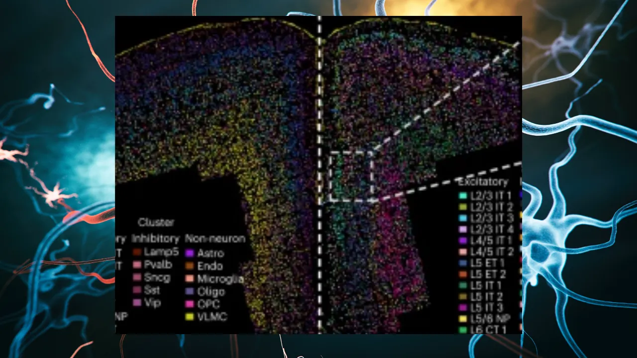
MERFISH (Multiplexed Error-Robust Fluorescence In Situ Hybridization) and Visium by 10x Genomics are both advanced techniques that enable spatial transcriptome analysis. MERFISH has high spatial resolution and can analyze gene expression at the single-cell level. Visium, on the other hand, can cover broad regions across an entire sample, but its spatial resolution is lower than that of MERFISH. Because the aim of this study was to classify cell types and subtypes in detail and to elucidate their spatial arrangement, MERFISH, with its high spatial resolution, was chosen.
Key Findings of the Study
Identification of Cell Types and Subtypes
In this groundbreaking study, the cell types and subtypes present in the mouse prefrontal cortex (PFC) were precisely identified. By using the advanced method of transcriptome analysis, the researchers were able to investigate the gene expression profile of each cell in detail and reveal the unique characteristics that each cell possesses. As a result, the diverse neurons and glial cells present in the PFC were identified, shedding light on the functional roles of each cell subtype.
Region-Specific Functions of Each Subregion
The PFC is divided into multiple subregions, each performing different functions. In this study, it was revealed that the distribution of cells differs across these subregions. For example, in the dorsal anterior cingulate cortex (dACC), L5 IT 3 neurons are mainly present, and this was suggested to be involved in the regulation of specific behaviors. In addition, in the prelimbic cortex (PL), L5 ET 1 neurons are abundant, and these were shown to contribute to the functions of the PL and the infralimbic cortex (ILA). These insights are extremely important for understanding the relationship between the region-specific functions of the PFC subregions and the cell types.
Cell-Cell Interactions and Their Effects
By identifying cell types and subtypes, the researchers were also able to deepen their understanding of how each cell interacts and transmits information within the brain. The networks and interactions among cells are essential for maintaining normal brain function, and this study revealed those complex relationships. This provided clues for understanding how abnormalities in neural circuits are involved in neurological diseases and cognitive impairments, and it is expected to contribute to the development of new therapies in the future.
Limitations of the Study and Future Prospects
Limitations of the Study and How to Overcome Them
Despite this innovative study, several limitations exist. First, the samples used were limited to the mouse prefrontal cortex, and other species, including humans, and other brain regions were not examined. Therefore, caution is needed in generalizing the insights obtained. In addition, the techniques used are highly advanced and generate large amounts of data, but their analysis requires specialized knowledge and resources. Furthermore, the interactions among cells and the dynamics of networks have not been fully elucidated, and further research is needed on how these are involved in the onset and progression of disease.
To overcome these limitations, additional studies targeting different species and brain regions are needed. The development of tools and algorithms for data analysis is also required. To understand cell-cell interactions and network dynamics, collecting data over time and combining it with other imaging techniques is effective.
Expectations and Potential for Future Research
By identifying the cell types and subtypes of the brain and revealing their spatial organization, this study opened up a new horizon in neuroscience. In future research, it is expected that, based on these insights, cell-cell interactions and network dynamics will be analyzed in even greater detail. This will allow a deeper understanding of brain function and disease mechanisms and contribute to the development of new therapies and preventive measures.
In addition, the techniques used in this study can be applied to other species, brain regions, and even different tissues and organs. This is expected to advance the understanding of cell types and functions across biology as a whole. Furthermore, by leveraging the large amount of data obtained in this study and using data analysis and machine learning techniques, it is also possible to contribute to the identification of new biomarkers and the early diagnosis of disease.
Overall, this study holds many possibilities not only in neuroscience but in biology as a whole, and its future development is highly anticipated.
Summary
Through this innovative study, we were able to deepen our understanding of the cell types and subtypes in the mouse prefrontal cortex and their spatial organization. This made it possible to unravel the brain’s complex networks and to take a major step forward in the field of neuroscience.
The Importance of the Study and Its Impact
The findings of this study are extremely important for understanding the relationship between the structure and function of the brain. In particular, by identifying cell types and subtypes and revealing how they are spatially organized, it becomes possible to analyze the mechanisms of brain function and disease in greater detail. This is expected to contribute to the early diagnosis of neurological diseases and the development of new therapies.
Contributions to Science and Medicine
This study holds the potential to have a major impact not only from the perspective of basic science but also in clinical practice. The detailed classification of cells and the understanding of their spatial organization contribute to the advancement of precision medicine and provide important information for delivering optimal treatment tailored to each individual patient.
In addition, the advanced techniques used in this study can be applied to other brain regions and different species, holding the potential to renew the entire field of brain science. This will further deepen our understanding of our own brains and bring a revolution to the medicine of the future.
Ultimately, this study contributes to unraveling the brain’s complex networks and opening new paths to improve treatments for neurological diseases. We look forward to the future possibilities this study brings and must continue to support the advancement of brain science.