Here is the paper we introduce this time.
This is a comparative evaluation of the storage conditions for extracellular vesicles.
In research and clinical settings, there is still no established method for how extracellular vesicle (EV) preparations should be stored to best maintain their therapeutic efficacy. This paper examines how the storage state affects EVs. Let us take a look.
Introduction:
The importance of extracellular vesicles (EVs) and their therapeutic applications
Extracellular vesicles (EVs) are minute particles secreted by cells that play a role in transmitting information between cells. They contain a diverse range of biomolecules that influence various physiological and pathological processes in the body, and are known in particular to carry proteins, ribonucleic acid (RNA), and even fragments of DNA. Owing to the diversity of their constituents and their role in intercellular communication, EVs have become an important subject of research in disease diagnosis, biomarker identification, and the development of novel therapies. In particular, they hold great promise for clinical applications such as cancer treatment, regenerative medicine, and immunomodulation. Therapies that harness these EVs are expected to maximize efficacy and minimize side effects by delivering specific therapeutic molecules directly to target cells.
Background and objectives of the study
The background to this study lies in the promise of EVs in therapeutic applications, together with the challenges that must be overcome to realize their clinical use. In particular, the impact of EV storage conditions on their quality and functionality is a crucial factor in maximizing therapeutic efficacy. This study aims to clarify the optimal conditions for the long-term storage of EVs and to evaluate strategies for maintaining their functionality. By comparing the changes in the physical and biochemical properties of EVs under different storage conditions and analyzing how these results affect the biological function and therapeutic efficacy of EVs, it seeks to provide important information toward the practical implementation of EV-based therapies. This study is expected to open the way to the development of new therapeutic strategies using EVs and to their clinical application.
Materials and methods used in this study
Cell culture, isolation and characterization of sEVs
In this study, small extracellular vesicles (sEVs) were isolated from a specific cell line, after which the properties of the sEVs were evaluated using various biological and physical methods. Cell culture used standard media, and the conditions for collecting sEVs from mature cells were optimized. Isolation was performed by methods such as ultracentrifugation and filtration, and the purity and yield of the obtained sEVs were evaluated.
Nanoparticle tracking analysis (NTA)
Nanoparticle tracking analysis (NTA) was used to measure the size distribution and particle number of the sEVs. This technique makes it possible to precisely evaluate the physical properties of sEVs and to understand the impact of the isolation process on sEV quality.
Evaluation of sEV cargo and study of cellular uptake
A detailed analysis of the biological molecules contained in sEVs, such as RNA and proteins, was carried out. The identification and quantification of these molecules are essential to understanding the biological function of sEVs. Furthermore, labeled sEVs were used to study the efficiency and distribution of cellular uptake, elucidating how sEVs act on target cells.
Biodistribution study
A biodistribution study was conducted to investigate the in vivo dynamics of sEVs. This involved administering labeled sEVs to an animal model and then tracking the distribution of the sEVs in tissues and organs. This study helps to evaluate the ability of sEV-based therapies to effectively reach the target tissue.
These methodologies provide a foundation for understanding in detail the impact of sEV storage conditions on their physical and biological properties.
Results
Characterization of sEVs
In this study, the properties of small extracellular vesicles (sEVs) under different storage conditions were evaluated. sEVs were isolated from the brain-derived endothelial cell line bEnd.3, and their size distribution and the presence of protein markers (CD63, TSG101, Alix) were evaluated by nanoparticle tracking analysis (NTA) and transmission electron microscopy (TEM). Fresh sEVs showed a consistent size distribution in NTA and TEM observations, and sEVs were still confirmed after storage at different temperatures. However, marked aggregation was observed after one week of storage 【14†source】.
Changes in sEV quantity and size
Further analysis of the sEV quantity after storage showed that the number of sEVs decreased rapidly under all storage conditions. Storage at -20°C and -80°C slowed the rate of decrease in nanoparticle number, but more than 40% of sEV particles were lost even after 28 days. Freeze-thawing also had a marked effect on sEV number. Freeze-thawing in liquid nitrogen caused severe damage to the sEVs, and freeze-thaw cycles between -20°C/-80°C and 4°C also contributed significantly to the loss of sEV particles.
Owing to the influence of storage conditions and freeze-thaw cycles, the relative quantity of sEVs differed; in particular, storage at -20°C notably increased the cumulative size distribution, with a loss of small particles (30-150 nm) and an increase in the proportion of large particles (150-500 nm) being observed. This indicates that the size range from D10 to D90 broadened under all storage conditions, and that storage at -20°C expanded the size more markedly.
These results revealed that different storage conditions have a marked effect on the quantity and size distribution of sEVs. In particular, it was shown that storage at -20°C increases the cumulative size distribution of sEVs, and that freeze-thaw cycles cause severe damage to the quantity of sEVs. These findings provide important considerations when selecting an sEV storage strategy.
Changes in cargo and cellular uptake
Storage conditions also affected the cellular uptake efficiency of sEVs. sEVs stored at 4°C showed a markedly reduced uptake efficiency by autologous cells, whereas sEVs stored at -80°C maintained a high uptake efficiency equivalent to the fresh state within three weeks. A decreasing trend was also seen in the uptake efficiency of sEVs stored at -20°C, but a marked difference emerged after 14 days of storage.
Changes in biodistribution
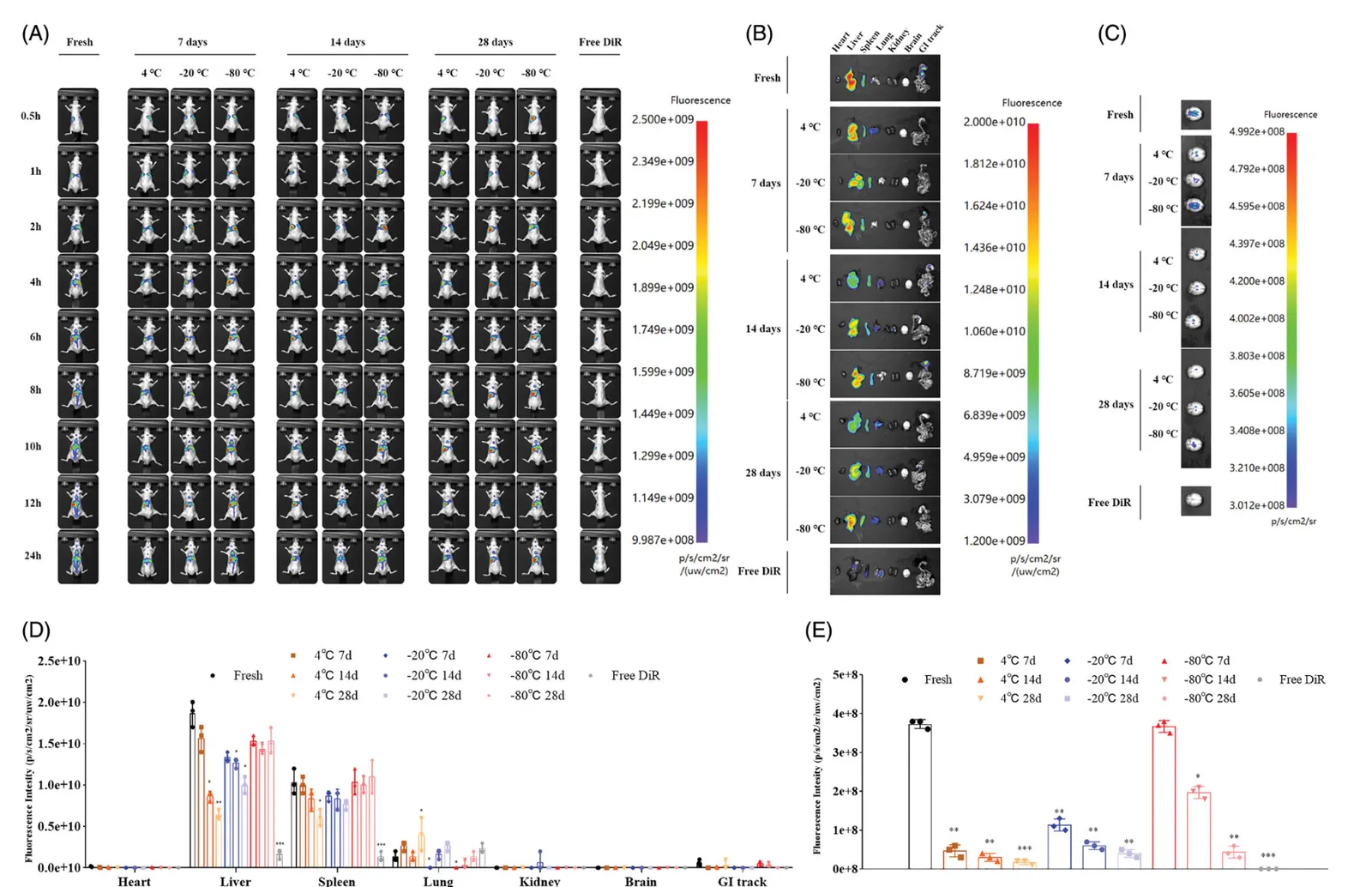
DiR-labeled fresh sEVs, or sEVs after storage, were administered to healthy mice by tail vein injection, and the biodistribution was imaged at different time points. Fresh sEVs showed a strong fluorescence signal throughout the body and especially in the brain, whereas in sEVs stored at 4°C or -20°C the fluorescence signal decreased markedly with the storage period. In particular, the fluorescence signal in the gastrointestinal tract and in the brain was barely detectable. On the other hand, in sEVs stored at -80°C, a stable fluorescence signal was observed in the mice and in dissected organs even during 28 days of storage. However, after 14 days of storage the fluorescence signal in the brain decreased markedly.
These results showed that the storage conditions of sEVs have a marked effect on changes in their cargo, on cellular uptake, and even on their distribution in vivo. In particular, they suggest that storage at -80°C best maintains the biological properties of sEVs, providing important information for the use of sEVs in therapeutic applications and as drug delivery systems.
Discussion
The impact of storage conditions on the stability and function of sEVs
This study revealed the impact of different storage conditions on the stability and function of extracellular vesicles (sEVs). In particular, storage at -80°C was shown to be optimal for preserving the quality of sEVs, which is consistent with the storage method recommended by the International Society for Extracellular Vesicles (ISEV). On the other hand, storage at 4°C and -20°C brought about marked changes in the size distribution, quantity, and cargo of sEVs, and these changes also affected the cellular uptake efficiency and biodistribution. These effects of storage conditions provide considerations for the use of sEVs in therapeutic applications and as drug delivery systems.
Significance of the study and future challenges
This study represents an important step toward advancing sEVs for clinical application. Understanding the impact of storage conditions on the function of sEVs lays the foundation for using these vesicles stably and effectively in therapy. However, the effects of different sEV sources and storage methods also need to be evaluated further, and the development of standardized storage protocols will be a challenge for future research. In addition, further research is needed to identify the optimal conditions for the storage of sEVs.
Conclusion
Key points of the study and outlook for clinical application
This study comprehensively evaluated the impact of sEV storage conditions on their size, quantity, cargo, cellular uptake efficiency, and biodistribution. Storage at -80°C was shown to best preserve the quality of sEVs, which is an important finding toward the clinical application of sEV-based therapies and drug delivery systems. Going forward, the development of detailed storage protocols and the exploration of new storage methods are encouraged in order to enhance the commercial availability of sEVs and to accelerate their translation to the clinic.