Lipid-associated macrophages linked to osimertinib resistance and leptomeningeal metastases in NSCLC
Lipid-associated macrophages for osimertinib resistance and leptomeningeal metastases in NSCLC
Lipid-associated macrophages linked to osimertinib resistance and leptomeningeal metastases in NSCLC
Journal name & year of publication
Cell Reports, 2024
First and last authors
Yang-Si Li, Yi-Long Wu
First affiliation
Guangdong Lung Cancer Institute, Guangdong Provincial People’s Hospital (Guangdong Academy of Medical Sciences), Southern Medical University, Guangzhou, China
Abstract
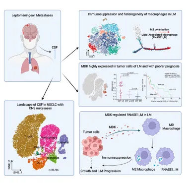
To clarify the role of lipid-associated macrophages in osimertinib resistance and leptomeningeal metastases (LM) in non-small cell lung cancer (NSCLC), single-cell RNA sequencing was performed on cerebrospinal fluid (CSF). In LM, heterogeneity of macrophages with immunosuppressive properties was confirmed, and a specific subset of lipid-associated macrophages (RNASE1_M) was shown to be involved in osimertinib resistance and the development of LM.
Background
Leptomeningeal metastasis in NSCLC is especially prominent after the emergence of osimertinib resistance, and treatment options for it are limited. In particular, immune evasion is thought to be a factor in osimertinib resistance, but the detailed mechanisms remain unclear.
Methods
Single-cell RNA sequencing was performed using cerebrospinal fluid from NSCLC patients harboring EGFR mutations. The study included patients with progressive disease and treatment-naïve patients, and characterization and functional analysis of cell clusters were carried out.
Results
The study confirmed that lipid-associated macrophages were increased in patients with LM, and it was revealed in particular that the RNASE1_M subtype is associated with osimertinib resistance. In addition, the MDK (Midkine) protein was shown to induce polarization of these macrophages.
Discussion
This study makes it possible to comprehensively understand the immune environment in leptomeningeal metastasis of NSCLC patients, and in particular it clarified the role of macrophages associated with osimertinib resistance. This suggests the possibility of new therapeutic targets.
Novelty compared with previous research
There is novelty in identifying lipid-associated macrophages in leptomeningeal metastasis and showing that they are associated with osimertinib resistance. In addition, the role of the MDK protein was newly identified.
Limitations
This study has limitations in that its sample size is small and it is restricted to cerebrospinal fluid samples. To address this, integration of existing data and validation in independent cohorts were carried out.
Potential applications
The RNASE1_M subtype and MDK protein identified in this study can be applied as future therapeutic targets and prognostic markers.
What is osimertinib?
Osimertinib is a third-generation EGFR tyrosine kinase inhibitor (TKI) used mainly to treat non-small cell lung cancer (NSCLC). It is particularly effective against lung cancers with EGFR gene mutations (such as exon 19 deletions and the L858R mutation), and it is also effective against tumors carrying the T790M mutation, which confers resistance to first- and second-generation TKIs.
Osimertinib binds to the mutant tyrosine kinase domain of EGFR and inhibits its activity, thereby suppressing the proliferation of cancer cells. It also has the ability to cross the blood-brain barrier and is sometimes used to treat central nervous system metastases (leptomeningeal metastasis and brain metastasis). Osimertinib is widely used as standard therapy, particularly for patients with advanced or metastatic EGFR-mutation-positive NSCLC.
What are lipid-associated macrophages (RNASE1_M)?
Lipid-associated macrophages (RNASE1_M) are a subtype of macrophage that highly express specific lipid-metabolism-related genes and are associated in particular with osimertinib resistance and the progression of leptomeningeal metastasis (LM) in non-small cell lung cancer (NSCLC). These macrophages have immunosuppressive properties and play a role in promoting immune evasion within the tumor microenvironment.
RNASE1_M macrophages highly express genes involved in lipid metabolism (for example, APOE and PLA2G7) and genes related to collagen degradation and the hypoxia response. Through this, they are thought to promote tumor growth and increase resistance to EGFR tyrosine kinase inhibitors such as osimertinib.
In addition, RNASE1_M macrophages have been shown to have their polarization induced by a protein called MDK (Midkine) secreted from tumor cells, and this interaction contributes to the formation of an immunosuppressive environment. Therefore, RNASE1_M macrophages are attracting attention for their potential as a new therapeutic target.
A comprehensive description of lipid-associated macrophages
Lipid-associated macrophages (LAMs) are a group of specialized macrophages closely linked to lipid metabolism, and they play important roles in a variety of pathological environments such as tumors, chronic inflammation, and metabolic diseases. These macrophages are present mainly in adipose tissue and lipid-rich environments and are involved in the uptake, metabolism, and storage of lipids.
Characteristics
-
Gene expression profile: LAMs highly express genes related to lipid metabolism. These include apolipoprotein E (APOE), fatty-acid-binding protein (FABP), glycosylated protein (LGALS3), and phospholipase (PLA2G7).
-
Function:
- Lipid uptake and storage: LAMs have the ability to take up lipids in serum and accumulate them internally. For this reason, their activity is especially high in lipid-rich locations such as adipose tissue, atherosclerotic plaques, and the tumor microenvironment.
- Immune regulation: LAMs often have immunosuppressive properties and, in the tumor microenvironment, play a role in promoting the immune evasion of tumor cells.
-
Pathological role:
- Tumor microenvironment: LAMs within tumors play a role in promoting tumor growth and progression and, in particular, exert immunosuppressive functions to help tumor cells escape from immune surveillance. For example, in non-small cell lung cancer (NSCLC) with EGFR mutations, LAMs have been reported to contribute to the acquisition of osimertinib resistance.
- Atherosclerosis: LAMs in atherosclerotic plaques affect plaque formation and stability and are involved in cardiovascular disease risk.
- Obesity and metabolic disease: In obese tissue, LAMs regulate the secretion of inflammatory cytokines and are involved in the development of insulin resistance and diabetes.
-
Regulatory factors: The function and phenotype of LAMs are regulated by factors secreted from tumor cells and other surrounding cells. For example, growth factors such as Midkine (MDK) have been shown to induce polarization of LAMs and promote tumor immune evasion.
Importance as a potential therapeutic target
Because LAMs play important roles in the regulation of the tumor microenvironment and chronic inflammation, new therapeutic strategies targeting these macrophages are being studied. In particular, it is hoped that controlling the activity of LAMs in combination with immune checkpoint inhibitors may strengthen the immune response against tumors.
A comprehensive understanding of LAMs may contribute to the development of new therapies in cancer treatment and chronic disease management.
How are LAMs related to neural regeneration?
The relationship between lipid-associated macrophages (LAMs) and neural regeneration is a field that has attracted attention in recent research. LAMs are macrophages involved in lipid metabolism and are mainly involved in the regulation of the tumor microenvironment and chronic inflammation, but they may also play important roles in the nervous system.
The relationship between LAMs and neural regeneration
- Lipid metabolism of the nervous system and LAMs: Nervous tissue, especially in the central nervous system (CNS), is rich in lipids, and lipid metabolism is essential for the function and health maintenance of nerve cells. LAMs are involved in the regulation of this lipid metabolism and may influence the process of neural regeneration. For example, it is conceivable that LAMs take up and process lipids around nerve cells and supply the energy and components needed for the regeneration and repair of nerve cells.
- Inflammation and neural regeneration: The inflammatory response after nerve injury plays a dual role in the regeneration process. On the one hand, excessive inflammation impedes neural regeneration, but moderate inflammation promotes clearance of the injured site and the regeneration process. LAMs may act as anti-inflammatory macrophages, supporting neural regeneration while controlling the inflammatory response.
- Regulation of supporting cells by LAMs: In the central nervous system, macrophages interact with microglia and astrocytes and play a role in preparing the environment needed for neural regeneration. LAMs can influence these supporting cells and directly or indirectly promote the regeneration of nerve cells.
- Clinical significance and research progress: The relationship between LAMs and neural regeneration is still a developing field of research, but in the future the development of new therapies targeting LAMs is anticipated in the treatment of nerve injury and neurodegenerative diseases. In particular, understanding how LAMs promote or inhibit neural regeneration may provide a foundation for devising more effective therapeutic strategies.
LAMs may play important roles not only in lipid metabolism but also in the process of neural regeneration, and they could be important regulatory factors in the mechanisms that promote regeneration and repair after nerve injury. Future research is expected to further clarify the specific relationship between LAMs and neural regeneration, as well as the possibilities of therapies utilizing this.
How are they related to M2 macrophages?
Lipid-associated macrophages (LAMs) are deeply related to M2 macrophages. M2 macrophages are known as anti-inflammatory macrophages and are involved in tissue repair, immunosuppression, and tumor progression, while LAMs similarly have immunosuppressive properties and are positioned as a subtype with characteristics specialized for lipid metabolism.
The relationship between M2 macrophages and LAMs
- Polarization and function: M2 macrophages are in an “alternatively activated” state that promotes inflammation suppression and tissue repair, and they secrete anti-inflammatory cytokines (such as IL-10 and TGF-β). LAMs, like M2 macrophages, also have an anti-inflammatory role and are particularly involved in the uptake and metabolism of lipids. These macrophages exert immunosuppressive effects in the tumor microenvironment and in sites of chronic inflammation, and they can help tumor growth and progression.
- Similarity of gene expression: LAMs and M2 macrophages often have a common gene expression profile. For example, CD206 (MRC1) and CD163, which are markers of M2 macrophages, may also be highly expressed in LAMs. This suggests that LAMs are a subset of M2 macrophages.
- Relationship with lipid metabolism: M2 macrophages are closely related to lipid metabolism. In environments where lipid metabolism is active, M2 macrophages more readily differentiate into LAMs, and the expression of lipid-metabolism-related genes (such as APOE and FABP5) in particular is enhanced. Through this, LAMs contribute to the progression of tumors and inflammatory diseases via lipid metabolism.
- Role in the tumor microenvironment: M2 macrophages play a role in promoting tumor immune evasion in the tumor microenvironment and promote tumor progression. Likewise, LAMs also have immunosuppressive effects in the tumor microenvironment and play a role in helping tumor cells escape from the immune system. In particular, in relation to osimertinib resistance in non-small cell lung cancer (NSCLC) with EGFR mutations, it has been shown that LAMs, by having the characteristics of M2 macrophages, promote treatment resistance.
LAMs are often classified as a subtype of M2 macrophages or as part of them, and the two are strongly related in that they are involved in immunosuppression, tissue repair, and lipid metabolism. In particular, in tumor and chronic inflammation environments, it is understood that M2 macrophages differentiate into LAMs, form an immunosuppressive environment via lipid metabolism, and contribute to disease progression.
What is the difference between M2 and LAM?
M2 macrophages and lipid-associated macrophages (LAMs) are both subtypes of macrophage that play immunosuppressive roles, but they differ in several respects. The main differences are shown below.
1. Definition and characteristics
-
M2 macrophages:
- Definition: M2 macrophages are “alternatively activated” macrophages involved in anti-inflammation and tissue repair, and they create an immunosuppressive environment.
- Characteristics: They secrete anti-inflammatory cytokines such as IL-10 and TGF-β, suppress inflammation, and promote tissue repair and regeneration. They are also known to be involved in parasitic infection, wound healing, and tumor progression.
-
Lipid-associated macrophages (LAMs):
- Definition: LAMs are a subtype of macrophage with characteristics related in particular to lipid metabolism, and they are activated in lipid-rich environments such as adipose tissue and the tumor microenvironment.
- Characteristics: They are specialized for the uptake and storage of lipids and highly express lipid-metabolism-related genes (such as APOE, FABP, and PLA2G7). They play immunosuppressive roles mainly in the tumor microenvironment and are involved in tumor progression and treatment resistance.
2. Gene expression and function
-
M2 macrophages:
- Gene expression: M2 macrophages highly express genes such as CD206 (MRC1), CD163, and Arginase-1 (ARG1).
- Function: They are involved in tissue repair, wound healing, parasite elimination, and promotion of tumor immune evasion. In general, they are involved in the resolution of inflammation and the control of chronic inflammation.
-
LAM:
- Gene expression: LAMs highly express lipid-metabolism-related genes such as APOE, FABP, LGALS3, and PLA2G7.
- Function: They are specialized for lipid metabolism and are involved in the uptake, accumulation, and metabolism of lipids. In particular, they play a role related to tumor growth and treatment resistance.
3. Tissue distribution and environment
-
M2 macrophages:
- Tissue distribution: They are found in various tissues throughout the body and are particularly activated at wound sites, sites of parasitic infection, and sites of chronic inflammation.
- Environment: They are often activated in repair processes after inflammation and in chronic inflammation environments.
-
LAM:
- Tissue distribution: They are present mainly in adipose tissue, lipid-rich environments, or the tumor microenvironment.
- Environment: They are activated in environments where lipid metabolism plays an important role, such as the tumor microenvironment, atherosclerotic plaques, and obese tissue.
4. Clinical significance
- M2 macrophages:
- Clinical significance: M2 macrophages promote wound healing and tissue repair, but they may also help tumor immune evasion and promote treatment resistance.
- LAM:
- Clinical significance: LAMs play a role in strengthening tumor growth and treatment resistance, especially in the tumor microenvironment, and are important in diseases related to lipid metabolism (such as atherosclerosis, obesity, and cancer). The potential of LAMs as a therapeutic target is being studied.
M2 macrophages and LAMs both have immunosuppressive properties, but LAMs differ in that they are particularly specialized for lipid metabolism. Whereas M2 macrophages have broader immune-regulatory functions and play roles in a variety of pathological situations, LAMs have roles specialized for lipid metabolism and the tumor environment. Understanding these differences is important in understanding disease pathology and in developing new therapies.
What kind of single-cell analysis is being performed?
Single-cell analysis is a technique for analyzing the gene expression profile of individual cells, and it is particularly useful for understanding highly heterogeneous cell populations. Regarding the single-cell analysis mentioned in the literature, the following techniques are used.
1. Single-cell RNA sequencing (scRNA-seq)
-
Purpose: By analyzing gene expression profiles at the single-cell level, it is performed to assess heterogeneity within cell populations and to clarify specific cell subtypes and their functional characteristics.
-
Target: Cells obtained from patients with non-small cell lung cancer (NSCLC) carrying EGFR mutations contained in cerebrospinal fluid (CSF).
-
Analysis procedure:
- Cells are separated and RNA sequencing is performed at the single-cell level to obtain gene expression data for each cell.
- Based on the obtained data, dimensionality reduction techniques such as t-SNE (t-distributed stochastic neighbor embedding) and UMAP (Uniform Manifold Approximation and Projection) are used to perform clustering of cell populations.
- For each cluster, the cell type is identified based on the expression of specific marker genes.
2. Characterization of cell clusters
- Identification of clusters:
- The obtained cell groups are classified into T cells, B cells, macrophages, monocytes, dendritic cells (DCs), epithelial cells, and so on, and further subtypes subdivided within these cells (for example, RNASE1_M and LYVE1_FOLR2_M) are identified.
- Functional scoring:
- The functional characteristics of each cell cluster (for example, antigen presentation, phagocytosis, and angiogenesis) are evaluated to clarify the role of each cluster.
3. Expression pathway analysis and trajectory analysis
-
Purpose: It is performed to trace the developmental trajectory of macrophages and to understand the formation process of subtypes associated with osimertinib resistance (such as RNASE1_M).
-
Method:
- A tree diagram showing the developmental trajectory of cells is created to visualize what pathways each cell follows.
- It is analyzed how the expression of specific genes changes along the trajectory, and genes and pathways associated with resistance are identified.
4. Analysis of cell-cell interactions
-
Purpose: To clarify what kind of interactions take place between macrophages and other cells (for example, tumor cells and T cells).
-
Method:
- Tools such as CellPhoneDB are used to predict ligand-receptor interactions between cells.
- Specific interactions (for example, the CD47-SIRPA pathway) are analyzed, and how they contribute to tumor immune evasion and treatment resistance is evaluated.
5. Identification of subtype-specific gene expression and regulatory factors
-
Purpose: To identify gene expression profiles characteristic of specific macrophage subtypes (for example, RNASE1_M) and to clarify the regulatory factors that control them.
-
Method:
- Genes showing differences for each subtype are identified, and their biological functions and the pathways in which they are involved are analyzed.
- In addition, regulatory networks of transcription factors are constructed, and it is clarified which transcription factors play important roles in specific subtypes.
In this study, diverse analytical techniques centered on single-cell RNA sequencing were used, and the heterogeneity of macrophages in the cerebrospinal fluid of non-small cell lung cancer and the mechanisms involved in treatment resistance were comprehensively analyzed. Through this, detailed understanding at the level of individual cells advances, and it is hoped that this will lead to the discovery of new therapeutic targets.
What instruments are used?
In this study, the following instruments and software were used to perform single-cell RNA sequencing (scRNA-seq):
Instruments
- BD Rhapsody Single-Cell Analysis System (BD, USA): Used for the preparation of single-cell libraries.
- Illumina NextSeq Platform: Used for sequencing.
- NanoDrop 2000 Spectrophotometer (Thermo Fisher Scientific): Used for quantifying RNA concentration.
- CFX96 Touch Real-Time PCR Detection System (Bio-RAD): Used for real-time PCR.
Software
- fastp: Used for filtering adapter sequences and removing low-quality reads.
- UMI-tools: Used for single-cell transcriptome analysis.
- STAR: Used for mapping to the human genome.
- Seurat: Used for normalization and clustering.
- Monocle 2.0: Used for reconstructing the developmental trajectory of macrophages.
- SCENIC: Used for transcription factor regulatory network analysis.
These instruments and software worked together to perform detailed gene expression analysis at the single-cell level, and the identification of macrophage subtypes associated with osimertinib resistance and leptomeningeal metastasis, the objective of the study, was carried out.