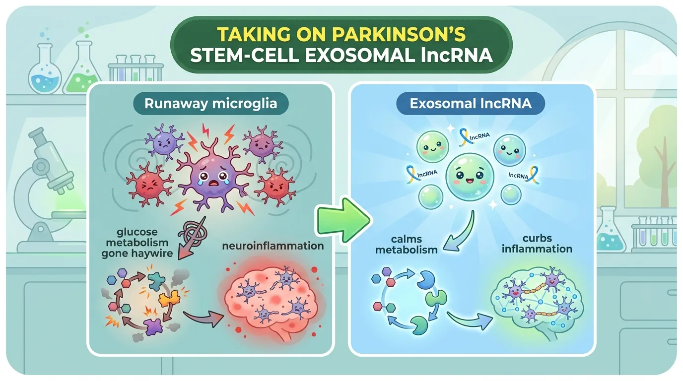
The hands tremble, the body stiffens, movements slow—Parkinson’s disease (PD) is the second most common neurodegenerative disorder after dementia. Its cause is the gradual death of dopamine neurons deep in the brain, in a region called the substantia nigra. Today’s treatments (levodopa and deep brain stimulation) ease the symptoms, but they cannot halt the underlying flow of neuron loss itself. That is precisely why researchers keep searching for new strategies to “stop the progression”—that is, to protect the neurons.
The paper I’m introducing today takes a slightly unexpected angle on that strategy: rewiring how the brain’s immune cells, “microglia,” use their fuel. The stars of the story are a tiny parcel released by stem cells taken from the mucosa deep in the nose—the exosome—and a single long non-coding RNA (lncRNA) packed inside it.
Journal Information
- Paper title: Olfactory Mucosal Mesenchymal Stem Cell-Derived Exosomal LncA2M-AS1 Ameliorates Parkinson’s Disease by Regulating Microglial Glucose Metabolic Reprogramming and Neuroinflammation via the CFL1/ROCK1 Axis
- Authors: Jiangshan Zhang (lead author), Guoshuai Yang, Yanhui Zhou, Dan Hou, Chuang Wang, Yujie Hu, Ying Xia (corresponding author)
- Affiliation: Department of Neurology / Neurosurgery, Haikou Hospital Affiliated to Xiangya School of Medicine, Central South University (Haikou, Hainan Province, China)
- Journal: CNS Neuroscience & Therapeutics (Wiley), 2026, Vol. 32, e71019
- DOI / Link: 10.1002/cns.71019
- Impact Factor: approx. 5.7 (2024 Journal Citation Reports, approximate. A well-regarded specialist journal ranked Q1 in the fields of neuroscience and neuropharmacology.)
- Open access: Yes (CC BY. Free to reuse and redistribute as long as the source is credited.)
About the corresponding author, Ying Xia: Ying Xia is a clinician-researcher in the Department of Neurosurgery at Haikou Hospital Affiliated to Xiangya School of Medicine, Central South University (Haikou People’s Hospital), and the senior (last) author of this paper. In a 2025 study that immediately preceded this one (Cell Biology and Toxicology), the same research group reported that lncA2M-AS1—carried by exosomes from the same olfactory mucosa-derived mesenchymal stem cells (OM-MSC)—teams up with a protein called IGF2BP1 to promote TP53INP1-dependent mitochondrial autophagy (the clearing-out of defective mitochondria), thereby easing oxidative stress in a Parkinson’s disease model. In other words, the present study is a sequel that digs into “how the same molecule works this time, from the angle of immunity and metabolism.” Across at least these two papers, the group can be positioned as one that consistently pursues the theme of tackling Parkinson’s disease with an lncRNA carried by OM-MSC exosomes, from a clinically grounded neurosurgical standpoint. (That said, as of this writing, what could be reliably confirmed from public information extends only to the affiliation, the role as corresponding author, and the research theme shared by these two papers. Because I could not pin down the Chinese-character rendering of the name, a detailed career history, or accomplishments in other fields, I have refrained from writing anything speculative here.)
👦 Student: Exotaro, what exactly was an “exosome” again?
🧬 Exotaro: It’s a tiny “parcel” that cells release into their surroundings, roughly 30–150 nanometers in diameter (one nanometer is a millionth of a millimeter). Packed inside are messages like proteins and RNA, ferried around like letters between cells. And because it’s made of a lipid membrane, it can reach the brain even across the “blood-brain barrier” (the checkpoint that guards the brain)—a barrier that drugs struggle to cross. Today’s star is a single RNA that was tucked inside one of those letters.
What Had Remained Unknown Until Now?
Parkinson’s Disease Is Not Only a Disease of “Dying Neurons”
Parkinson’s disease may be strongly pictured as a disease in which dopamine neurons dwindle. But what has become clear in recent years is that neuroinflammation plays a major role as an “accomplice” that eggs on the death of those neurons.
The cells that stand watch over immunity inside the brain are called microglia. Normally they patrol the brain as janitors, clearing away debris and foreign matter—but in Parkinson’s disease they become excessively activated. When that happens, they scatter inflammation-causing substances (cytokines), injure dopamine neurons, and even damage the gate that protects the brain (the blood-brain barrier). In short, the “protector” turns into a “rioter.”
👦 Student: Why would a janitor start running wild?
🧬 Exotaro: The key lies in a change in how the cell “uses its fuel.” That’s the most fascinating part of this paper.
Microglia in “Runaway Mode” Guzzle Sugar
Our cells normally use a fuel-efficient way of generating power called oxidative phosphorylation, which extracts energy (ATP) efficiently from sugar and fat. But once microglia are activated and enter combat readiness, they switch this over to a different method called glycolysis. Glycolysis is like a “fast charge” that rapidly breaks down sugar without using oxygen; it can produce energy quickly, but it is fuel-inefficient and stockpiles large amounts of “combustion residue” such as lactate and reactive oxygen species (ROS).
This metabolic switch closely resembles the “Warburg effect” seen in cancer cells, and it is deeply tied to the “transformation switch” that turns microglia into their aggressive, inflammation-causing (M1-like) state. And the accumulated lactate and ROS stoke yet more inflammation—a vicious cycle.
From here, an idea takes shape: “If we could rewire microglial glucose metabolism from ‘combat mode’ back to ‘normal mode,’ might we be able to calm neuroinflammation?” This very notion of metabolic reprogramming was the starting point of the present study.
The “Deep-Nasal Stem Cell” Pinned as a Therapeutic Courier
So how do you rewire microglial metabolism? The courier the authors chose is the olfactory mucosa-derived mesenchymal stem cell (OM-MSC).
Mesenchymal stem cells (MSC) are well known as a mainstay of regenerative medicine, and OM-MSCs in particular have advantages. They are obtained from a comparatively easy-to-access site—the mucosa deep in the nose—can potentially use the patient’s own cells (autologous transplantation), and carry the aptitude to grow into nervous-system cells. And crucially, much of the therapeutic effect of MSCs is delivered not by the cells themselves but by the exosomes the cells secrete. Exosomes load up “message substances” such as proteins, microRNA (miRNA), and lncRNA, and hand information from cell to cell.
The authors’ earlier work had established that OM-MSC exosomes are richly loaded with a long non-coding RNA called lncA2M-AS1. An lncRNA does not itself serve as a blueprint for protein, yet it is a “command-tower”-like molecule that fine-tunes how genes work. But how this lncA2M-AS1 manipulates the immunity and metabolism of microglia had not yet been unraveled. This is the gap the present study set out to fill.
The Clues: Two Molecules Called “CFL1” and “ROCK1”
The authors obtained one promising clue from computational analysis (bioinformatics): lncA2M-AS1 looked likely to bind the blueprint (mRNA) of a protein called CFL1 (Cofilin 1). CFL1 works to break down the cell’s scaffolding (actin), and in Parkinson’s disease it was known to promote the aggregation of α-synuclein and the impairment of neurons.
Furthermore, CFL1 was connected to another important protein, ROCK1 (Rho-associated coiled-coil containing protein kinase 1). ROCK1 is a “field unit” that carries out the cell’s various orders, and it is involved in switching glucose metabolism (glycolysis). On top of that, ROCK1 had been reported to trigger abnormal fragmentation of mitochondria through a molecule called Drp1, promoting the death of dopamine neurons.
Here the authors put forward a bold hypothesis: “Through a single communication line—lncA2M-AS1 → CFL1 → ROCK1—this stem-cell exosome may be controlling the metabolism and inflammation of microglia.” The present study tested this hypothesis in cells, in mice, and in human serum.
What Did This Paper Find?
To state the conclusion first: the authors’ hypothesis was largely borne out. Let’s walk through it step by step.
1. In Mice: The Exosomes Protected Motor Function and Calmed Brain Inflammation
First, using mice made parkinsonian with a neurotoxin called MPTP (20 mg/kg per day for 14 days), the researchers injected OM-MSC exosomes into the cerebral ventricles. The result—
- Motor function improved: In the open-field test, the distance traveled increased, movement speed rose, and time spent immobile decreased. The abnormal rotational movement induced by apomorphine also declined.
- Neurons were protected and inflammation subsided: When the substantia nigra was stained and examined, TH (tyrosine hydroxylase)—a marker of dopamine neurons—increased (= neurons preserved), while IBA1, a marker of activated microglia, decreased (= inflammation quieted).
- The brakes went on glycolysis: Four proteins involved in glycolysis—GLUT1 (the sugar-uptake port), HK2, PKM2, and LDHA—all dropped, and inflammatory cytokines (TNF-α, IL-1β, IL-6) fell as well.
The clincher was a “subtraction experiment.” When lncA2M-AS1 was removed (knocked down) from the exosomes, all of these beneficial effects weakened together (for the mice’s motor function, the improvement was partially canceled, returning to roughly midway between the untreated and the normal state). In other words, what was working was not so much the exosome itself as the lncA2M-AS1 loaded inside it.
👦 Student: So if it stopped working once you removed the exosome’s contents, that means…?
🧬 Exotaro: Right—it becomes strong evidence that “this RNA is the true identity of the effect.” They didn’t just show it was “a parcel with something good inside”; they pinned down “which letter did the work.”
2. In Cultured Cells: The Microglial “Runaway Metabolism” Was Directly Calmed
Next, the test-tube experiments. When the mouse microglial cell line BV2 was pushed into an inflammatory state by adding LPS (a bacterial toxin component), the cells weakened and tilted heavily toward glycolysis. Adding OM-MSC exosomes here—
- Cell viability recovered
- ECAR (the extracellular acidification rate, an index reflecting the momentum of glycolysis) fell, putting the brakes on runaway glycolysis
- OCR (the oxygen consumption rate, an index of mitochondrial respiration) recovered, and the fuel-efficient mode of power generation returned
- Lactate decreased, and the glycolytic proteins (GLUT1, HK2, PKM2, LDHA) dropped as well
Fluorescently labeled exosomes were properly taken up by BV2 microglia about three hours after being added. And here too, when lncA2M-AS1 was knocked down, these effects vanished. The phenomenon seen in mice was reproduced at the cellular level along the very same storyline.
Even more ingenious is the “co-culture” experiment of microglia and neurons. When injured microglia (BV2) and neurons (HT22) were grown together in the same environment, the neurons weakened and inflammatory substances increased. Yet when they were grown together with microglia that had been pretreated with OM-MSC exosomes, the neurons’ viability recovered and the inflammation subsided as well. “Calming the microglia ends up protecting the neurons”—this connection was demonstrated experimentally.
3. In Human Serum: The Molecules’ “Ups and Downs” Were Tied to the Disease
The authors also examined serum from 15 Parkinson’s disease patients (8 men, 7 women; mean age 68.5 ± 6.2) and 15 age- and sex-matched healthy controls. The result—
- lncA2M-AS1 was decreased in the patients
- Conversely, ROCK1 was elevated in the patients, and the two showed a negative correlation (when one is high, the other is low; P=0.0253, R²=0.3293)
When lncA2M-AS1 falls, ROCK1 rises. In terms of direction, this relationship agrees with the hypothesis that “lncA2M-AS1 acts as a brake that suppresses ROCK1” (as discussed later, the correlation is only moderate in strength, but the direction is as the hypothesis predicts).
4. The Core Mechanism: The lncRNA Cuts Down the “Bodyguard” to Get ROCK1 Eliminated
Now for the heart of the matter: the molecular machinery. This is the most intricate part of the study and slightly counterintuitive, so let’s follow it carefully.
First, a luciferase reporter assay confirmed that lncA2M-AS1 attaches directly to the mRNA of CFL1. When lncA2M-AS1 was increased, the luminescence (= the momentum of CFL1 translation) dropped by about half in the normal sequence (CFL1-WT), while it did not change in the sequence with the binding site destroyed (CFL1-MUT). In other words, lncA2M-AS1 selectively suppresses the “reading-out” of the CFL1 blueprint. Indeed, in patient serum CFL1 was high (Fig 4A) and again showed a negative correlation with lncA2M-AS1 (P=0.0224, R²=0.3404).
So why does ROCK1 also fall when CFL1 decreases? This is the crux. CFL1 was working as a “bodyguard” that protects the protein ROCK1 from degradation. Cells have a mechanism that tags no-longer-needed proteins with a marker called “ubiquitin” and sends them to the garbage-disposal plant (the proteasome). By getting in the way of this marker being attached to ROCK1, CFL1 was keeping ROCK1 alive longer.
The story therefore connects like this:
- lncA2M-AS1 increases →
- CFL1 translation is suppressed, and CFL1 decreases →
- The bodyguard protecting ROCK1 is gone, so ubiquitin attaches to ROCK1 and its degradation proceeds →
- ROCK1 decreases
In the experiments, too, increasing lncA2M-AS1 reduced both the CFL1 and ROCK1 proteins, increased the ubiquitination of ROCK1, and also lowered the stability of ROCK1 mRNA. And when CFL1 was supplemented at the same time, these changes were pushed back (though not completely). This confirms that it was precisely CFL1 that held ROCK1’s fate in its hands.
👦 Student: “Reduce CFL1 and ROCK1 falls too”… normally I’d think reducing something just makes it go down, plainly enough.
🧬 Exotaro: Good point to press on. This is where it’s counterintuitive. CFL1 was ROCK1’s “bodyguard.” So when you cut down the bodyguard (CFL1), the one being protected (ROCK1) gets sent off to disposal and dwindles. lncA2M-AS1 wasn’t hitting ROCK1 directly; it was eliminating it indirectly by removing its bodyguard—that’s the idea.
5. The Storyline Held Up Even in the Reverse Experiments
The authors took further care. Directly knocking down CFL1 again reduced ROCK1 and calmed both microglial glycolysis and inflammation. But when ROCK1 was forcibly increased there, that beneficial effect was canceled out. In the same way, the benefit obtained by increasing lncA2M-AS1 was also nullified when ROCK1 was increased. The storyline that “the effect hinges on reducing ROCK1” was confirmed from every angle.
Finally, when lncA2M-AS1 was overexpressed in the mice’s bodies using AAV (adeno-associated virus), brain CFL1 and ROCK1 went down, motor function improved, the neurons seen by Nissl staining were preserved, TH increased, and IBA1, the glycolytic proteins, and inflammatory cytokines went down. From the culture dish to the living mouse, a single communication line—lncA2M-AS1 → CFL1 → ROCK1 → microglial metabolism → neuroinflammation—was consistently borne out.
How Will the Future Change? (The Road to the Clinic)
The significance of this study lies in having sketched out, in molecular terms, a new therapeutic angle on Parkinson’s disease: “calming neuroinflammation from upstream, at the level of microglial metabolism.” What’s more, its appeal is that the means employs a next-generation courier—“loading the lncRNA that is the true agent of the effect onto exosomes released by stem cells, and delivering it.”
Several concrete leads also came into view for thinking about clinical application.
- Potential as a biomarker (a diagnostic marker): The fact that the serum “ups and downs” of lncA2M-AS1, ROCK1, and CFL1 divided clearly between patients and healthy people suggests these could, in the future, become clues for telling by blood whether someone has Parkinson’s disease. (That said, this study did not verify whether these molecules also reflect the severity or the degree of progression of the disease, so that remains a task for the future.)
- Refining the delivery method: In this study the exosomes were delivered by direct injection into the cerebral ventricles (ICV administration). While this reliably reaches the brain, it is a surgical method with a large burden on the body. The authors mention the possibility of a gentler intranasal administration (delivery through the nose). Given that OM-MSCs are themselves derived from the mucosa deep in the nose, this “from the nose to the brain” route seems well matched, and its verification is anticipated going forward.
But—here, as a physician, let me state this clearly. All of the present findings are results at the “preclinical” stage. That is, they rest on experiments in mice with an MPTP-induced Parkinson’s disease model and in cultured cells (BV2 microglia and HT22 neurons). The data obtained in humans amount only to the observation that the amounts of the molecules in serum correlated with the disease; we are by no means at the stage of having given this treatment to people and shown it worked or was safe. Before clinical application, a long road remains: verification in larger animals, safety testing, and careful clinical trials in humans.
Even so, the direction is highly promising. The idea of “gently rewiring the disease-disrupted ‘metabolic settings’ of immune cells with a stem-cell-derived lncRNA” holds the potential to extend beyond Parkinson’s disease to many brain diseases in which neuroinflammation is involved.
How to Read This Study Critically—Its Limitations and the Path to Even Higher Quality
A good reader never swallows results whole; they always ask, “How certain is this?” As Exotaro, let me hold this paper to the same measuring stick. To be fair first: the study’s design for pinning down causation is solid. The “subtraction (knockdown)” that narrows the true agent of the effect down to the single lncA2M-AS1, the “reverse verification” in which adding ROCK1 back makes the effect disappear, and the corroboration from three directions—mice, cultured cells, and human serum—these deserve high marks. On that basis, let’s look honestly at the limitations as well.
Limitation 1: The model does not fully mirror human Parkinson’s disease. MPTP is a standard model that acutely injures dopamine neurons with a neurotoxin, but it does not reproduce the α-synuclein aggregation (Lewy pathology) at the core of human Parkinson’s disease, nor the chronic course that unfolds over many years. On the cell side, too, the microglia are BV2 and the neurons are HT22 (a hippocampus-derived cell line)—not human substantia nigra dopamine neurons themselves. The authors themselves acknowledge that HT22 does not adequately represent substantia nigra dopamine neurons. → What could have been done better: If the findings could be reproduced in a chronic model using α-synuclein fibrils or genetic modification (e.g., A53T), or in human iPSC-derived midbrain dopamine neurons and human microglia, the reliability of extrapolation to humans would rise a notch.
Limitation 2: The human data amount only to “a small-scale serum correlation.” The sole human evidence is the up-and-down changes and correlations of lncA2M-AS1, ROCK1, and CFL1 in the serum of 15 patients and 15 healthy people. The correlations are all moderate (the coefficient of determination R² is around 0.33, meaning it explains only about 30% of the variance), and blood does not directly mirror the state of microglia and neurons inside the brain. → What could have been done better: With larger numbers, ideally also measuring cerebrospinal fluid (CSF), and under a pre-registered analysis plan, evaluating “whether it can predict diagnosis or disease stage” with numbers like sensitivity, specificity, and AUC would make its true worth as a biomarker “auditable.”
Limitation 3: There are still unresolved seams in the central mechanism. The claim that “lncA2M-AS1 suppresses CFL1 translation” does not yet specify which part of the CFL1 mRNA it binds to, and how (the authors likewise leave this as future work). Furthermore, although the main thread is that “the CFL1 protein protects the ROCK1 protein from degradation (= post-translational stabilization),” in the experiments lncA2M-AS1 also lowered the stability of ROCK1 mRNA (Fig 4H). A protein-level account and an mRNA-level account coexist here, and the relationship between the two is not fully sorted out. In addition, exactly how CFL1 blocks the ubiquitination of ROCK1 is, in part, discussed by analogy from a precedent with a different molecule (PLD1). → What could have been done better: Directly demonstrate the substance of the binding via RNA pulldown or introduction of mutations at the binding site, and verify by separating out the three—“translational suppression / mRNA stability / protein degradation.” Further confirming that a ROCK1-selective inhibitor yields the same result would tighten the causal logic of the pathway.
Limitation 4: Room for improvement in delivery, safety, and the thoroughness of reporting. In this study the administration was intracerebroventricular injection (ICV), the animal experiments used roughly six mice per group, and the western blot quantification was on the order of three samples—the scale is on the small side. Overexpression via AAV is also forced expression beyond the physiological range. The observations, too, centered on motor function at 4–6 weeks after administration, and did not press on to long-term safety or toxicity, or to comparisons by route (ICV vs. intranasal vs. intravenous). In addition, procedures such as randomization and blinding of the behavioral tests, and the handling of multiple comparisons, are hard to read off from the main text. Note that there is a passage in the text stating that CFL1 is “decreased in PD,” but this conflicts with the rise in CFL1 shown by the figure (Fig 4A) and with the mechanism of the paper as a whole (lncA2M-AS1↓ → CFL1↑ → ROCK1↑). In this article I have read it, in line with the figure and the mechanism, as “CFL1 is increased in PD”—whether you can notice such “wobble” in the original’s wording is also part of reading critically. → What could have been done better: Adding a pre-registered animal-experiment protocol plus randomization, blinding, and multiple-comparison correction; route-by-route biodistribution and dose-finding; and long-term safety evaluation would make the bridge to the clinic far more realistic.
On the whole, none of this negates the paper’s value. The conception of “realigning neuroinflammation from one step upstream, at microglial glucose metabolism, with a stem-cell-derived lncRNA,” and the design that carefully traced its causation, are genuinely a step forward. But to be honest—this is a “proof of promising preclinical concept,” not a “proof of efficacy in humans.” Its true worth will be tested by the verification that lies ahead, in more genuine models and in humans.
Exotaro’s Perspective
This paper connects straight to my own research theme, and I found myself nodding again and again as I read. I have worked on research that puts extracellular vesicles (EV) / exosomes derived from mesenchymal stem cells (MSC) to use in treating neurological disorders, beginning with spinal cord injury (SCI). The cell source differs (today’s star is the olfactory-mucosa-derived OM-MSC, harvested from a different site than the MSCs I work with), but the foundation—“using MSC exosomes as a therapeutic platform for neurological disease”—is exactly what I face day to day.
What moved me most was the perspective of “switching the face and the metabolism of microglia.” In the setting of spinal cord injury as well, whether the immune cells at the injury site (microglia and macrophages) switch from an aggressive, inflammation-stoking state to a gentle, repair-assisting state greatly determines recovery. This paper laid its hand on the lever that lies deep behind that “switch”—energy metabolism (glycolysis vs. oxidative phosphorylation)—and, moreover, showed it being moved with a single lncRNA. Not “calming” immunity, but “realigning” it from its underlying metabolism—this idea is sure to become ever more important in the field of neural regeneration as well.
Another thing I learned much from as a researcher is the tenacity toward “how far to pin down the true agent of the effect.” An exosome is like a “grab bag” loaded with hundreds of kinds of molecules, and it is not hard to merely say “it worked.” But this study inserted and removed the lncA2M-AS1 inside, verified its downstream CFL1 and ROCK1 one by one, and drew the causal chain all the way to “this single RNA works through this pathway.” If you aim for a drug to carry into the clinic, it is precisely this resolution of “why it works” that becomes the lifeline.
As someone aiming for the same horizon—carrying regenerative medicine to the patient’s bedside—this was a paper that greatly inspired me. That the day may come when a parcel released by tiny stem cells deep in the nose gently calms inflammation in the brain—as one who walks the same road, I hope for it from the bottom of my heart.