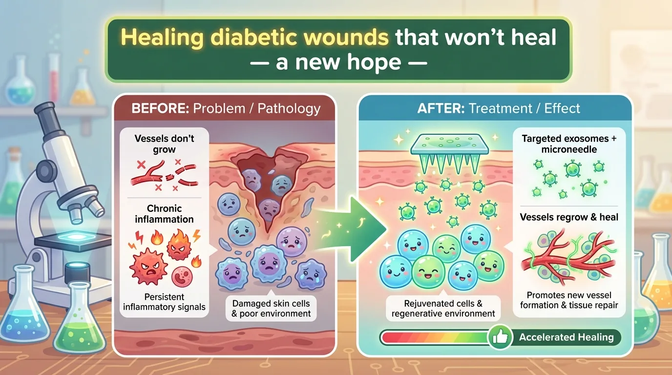
Among the complications of diabetes, the one that torments patients and clinicians the longest is the diabetic wound, epitomized by the “foot ulcer.” A small wound that would close in a few days in a healthy person goes weeks or months without healing in diabetes, and sometimes progresses to infection or even amputation. What I introduce today is a paper that confronts head-on the true nature of that “failure to heal.” An exosome — a cell’s little parcel — given the power to summon blood vessels back, delivered deep into the wound by a “dissolving microneedle (MN)” finer than a strand of hair: this SF-like treatment strategy achieved a solid result in diabetic mice.
Journal Information
- Paper title: Breaking the vicious cycle of impaired angiogenesis and chronic inflammation in diabetic wounds: a bioengineered microneedle system delivering RGD-modified M2 exosomes
- Authors: HongYu Wang, BaoHua Wei, BaiShi Wang, Mi Chai, Jing Ren, Yan Han, LingLi Guo (Department of Plastic and Reconstructive Surgery, Chinese PLA General Hospital, and others)
- Journal: Burns & Trauma (Oxford University Press), 2026, Vol. 14, tkag013
- DOI / link: 10.1093/burnst/tkag013
- Impact Factor: approximately 9.8 (2024 Journal Citation Reports, estimate. This is a high-impact journal in the burns and wound field, which has been trending upward in recent years.)
- Open access: Yes (CC BY-NC 4.0. As long as it is non-commercial, you are free to reuse it with attribution.)
👦 Student: Exotaro, “exosome” came up before too, but what was it again, exactly?
🧬 Exotaro: It’s a tiny “parcel” that cells put out, 50–150 nanometers in diameter (a nanometer is one-millionth of a millimeter). Packed inside are messages like proteins and microRNA (miRNA), exchanged like “letters” between one cell and another. Today’s star is the story of taking this parcel and remodeling it by hand into a “healing drug.”
What Did We Not Understand (or Fail to Achieve) Until Now?
The “Two Reasons” a Diabetic Wound Won’t Heal
By estimates from the International Diabetes Federation (IDF), the diabetic population will reach 643 million by 2030 (an adult prevalence of 11.3%) and 783 million by 2045 (12.2%). At least 5–10% of them will experience the complication of a diabetic wound. These are by no means someone else’s numbers.
So why don’t wounds heal in diabetes? The core point the paper makes is that two pathological conditions are occurring at the same time.
- Impaired angiogenesis — To heal a wound, new blood vessels that carry oxygen and nutrients must extend to the site. In diabetes, however, this “power to build new blood vessels” is weakened. If you cannot lay water pipes to a plot of land, no house can be built on it. In the same way, without the supply route that blood vessels provide, tissue cannot regenerate.
- Chronic inflammation — Inflammation is supposed to flare up briefly in the early phase of healing and then finish its job. In a diabetic wound, however, the inflammation does not subside and drags on and on.
The troublesome part is that these two form a “vicious cycle” in which they drag each other down. When blood vessels are scarce, tissue becomes starved of oxygen, and that fans inflammation. When inflammation persists, the environment for building blood vessels grows even more hostile — throughout this paper, the authors set as their theme the goal of “severing this vicious cycle itself” (this is the authors’ claim and hypothesis, and here they set out to test it in mice).
Let’s look a little more at the cellular level. When high blood sugar persists, AGEs (advanced glycation end products) — formed when sugar in the blood binds to proteins — increase and batter the endothelial cells that line the inside of blood vessels. You could rephrase it as sugar’s “scorching” rusting the blood vessels. In this paper, too, in vascular endothelial cells injured by high glucose and AGEs, proteins that tilt toward inflammation and cell death (ICAM-1, NF-κB, Bax) increased, the protein that protects cells (Bcl-2) decreased, apoptosis (a cell’s self-destruction) actually rose, and their power to migrate dropped substantially. The very cells that are the protagonists of blood-vessel building are weakened — this lies at the foundation of why diabetic wounds do not heal.
👦 Student: Then couldn’t you just slather on a drug that grows blood vessels?
🧬 Exotaro: You’ve hit on a good point. In fact, the star player for growing blood vessels is a protein called bFGF (basic fibroblast growth factor). But this one is a tricky customer.
The “Three Weaknesses” of Existing Treatments
bFGF, which grows blood vessels, is an attractive candidate, but using it on a wound as-is has the following weaknesses.
- Fragile: bFGF is unstable and has a short half-life, breaking down quickly in the environment of a wound. Its effect vanishes before it arrives — a “drug of easily melting ice,” so to speak.
- Doesn’t reach deep: Conventional topical agents such as gels and ointments tend to leave the drug at the “surface” of the wound. Yet regeneration and immune regulation take place in the deeper dermis layer. Merely applying to the surface fails to spread the drug to the very site that matters.
- Can attack only one problem: And the biggest weakness is that it cannot take on the two pathological conditions — angiogenesis and chronic inflammation — at the same time. Even if you fix one, the other keeps the vicious cycle spinning.
In other words, what has been missing until now is a treatment that is “unbreakable, reaches deep, and can strike both problems at once.” This paper set out to pack all three beats into a single system.
What This Paper Reveals
The authors’ answer was a combination: deliver “engineered exosomes” with a “dissolving needle.” Let’s walk through their intricate design step by step.
Step 1: Remodeling Macrophages into a “Drug Factory”
First, they chose a macrophage (the cell that serves as the immune system’s cleanup crew and command center) cell line (RAW264.7). A macrophage has, broadly, two faces.
- M1 type: the “attacking face” that stirs up inflammation (markers = CD80, CD86, iNOS)
- M2 type: the “healing face” that calms inflammation and repairs tissue (markers = CD206, CD163, Arg-1)
Into this macrophage the authors inserted the bFGF gene using a lentivirus, creating a stable cell line that keeps producing bFGF in large amounts (overexpression). They then added IL-4 (interleukin-4) and IL-10 at 20 ng/mL each for 36 hours to differentiate the cells toward the M2 type, the “healing face” (M2 polarization). What matters is that even after becoming M2, the cells’ power to make plenty of bFGF did not decline. In this way a parent cell was produced that takes both the “inflammation-calming property (M2-derived)” and the “power to grow blood vessels (bFGF).”
From these, exosomes are collected by ultracentrifugation (100,000×g). What you get at this point is called E-Exos (engineered exosomes).
👦 Student: Why make them from macrophages rather than from mesenchymal stem cells (MSCs)?
🧬 Exotaro: Sharp question. Macrophage-derived exosomes have a “homecoming” property — remarkably, they are readily taken up by macrophages themselves. So the villainous M1 macrophages present at the wound can be efficiently “persuaded” back into virtuous M2 ones. This exploits the general tendency for exosomes to reach the cell that “gave birth” to them.
There is a reason exosomes excel as “carriers” in the first place. Because they are made of the same lipids as the cell membrane, they can fuse membrane-to-membrane with the target cell, be swallowed up (endocytosis), or bind to receptors like a key in a lock, gently handing their contents — proteins, nucleic acids, and lipids — into the interior of the cell. Unlike synthetic nanoparticles, they are not easily disliked by the immune system, are low in toxicity, and are stable — which is exactly why they are called “next-generation carriers.” And their action mirrors the individuality of the cell that “gave birth” to them. Harvest them from M2 macrophages and you get “letters that calm inflammation”; harvest them from cells loaded with bFGF and you get “letters that grow blood vessels.” The TE-Exos of this study are a greedy parcel that seals both into a single letter.
Step 2: Taking Aim at Blood Vessels with the RGD Peptide
The engineered exosomes still had a weakness: their power to gather at the intended site (targeting) is weak, and they vanish quickly inside the body. You can write the letter, but with a vague address it is prone to going astray in the mail.
So the authors attached the RGD peptide (arginine-glycine-aspartic acid, a three-amino-acid sequence) to the surface of the exosomes. RGD binds tightly to integrin αvβ3, a receptor that appears abundantly on the surface of vascular endothelial cells activated to heal a wound. In other words, RGD is an address label marking the “construction site for new blood vessels.”
This receptor αvβ3 is a truly well-made “target.” On vascular endothelial cells, which are usually quiet, it barely shows its face; but the moment the cells receive stimuli such as bFGF, VEGFR (vascular endothelial growth factor receptor), or IL-8 and begin building new blood vessels, it suddenly increases on the surface. Moreover, at the front line of new blood vessels it activates MMP-2 (matrix metalloproteinase-2), “leveling the ground” of the surroundings to help endothelial cells migrate. Put another way, αvβ3 is like a flag that flies only on “cells that are building blood vessels right now.” Aim at that flag with RGD and you can concentrate the drug at the site that needs healing. It is also known that the cyclic peptide (cRGDfk) used here binds more strongly to blood vessels than does linear RGD.
For the attachment, they use a molecule called DSPE-PEG-cRGDfk-FITC. The lipid at one end (DSPE) anchors into the exosome membrane like an anchor and holds fast, while the RGD at the other end (the cyclic peptide cRGDfk) points to the blood-vessel address — a clever design that applies an “address sticker” without breaking the exosome’s structure. What is completed in this way is the star of this paper, TE-Exos (Targeted Engineered Exosomes).
Under transmission electron microscopy (TEM), both E-Exos and TE-Exos were confirmed as round (cup-shaped) particles 50–150 nm in diameter, and the exosome marker proteins CD9 and CD81, along with the cargo bFGF, were detected. As intended, TE-Exos were taken up in greater numbers by vascular endothelial cells injured by high glucose. The address sticker worked exactly as planned.
Step 3: Reinvigorating Injured Blood-Vessel Cells (in the Test Tube)
Next, they gave exosomes to cells that mimic diabetic blood vessels — human umbilical vein endothelial cells (HUVEC) injured by high glucose (treated with 20 mM glucose and 100 μg/mL AGEs for 48 hours) — and observed the response. The results were clear-cut.
- Proliferation (EdU and CCK-8 assays): Both E-Exos and TE-Exos increased cell numbers, but TE-Exos were the most potent of all. At 48 and 72 hours of culture, TE-Exos significantly outperformed E-Exos.
- Migration (scratch assay): TE-Exos also gave the cells the highest “power to migrate.” To close a wound, cells must move and gather, so this ties directly to healing.
- Tube formation (the power to build the tubes that become blood vessels): The TE-Exos group had the greatest number of meshes (network loops), indicating a high power to assemble blood vessels.
Furthermore, in experiments using mouse macrophages (peritoneal macrophages and bone marrow-derived macrophages), adding TE-Exos decreased the attacking M1 markers (CD80, CD86) and increased the healing M2 markers (CD206, CD163), lowering the M1/M2 ratio — that is, the immune mood shifted from inflammation-leaning to repair-leaning. The power to grow blood vessels and the power to calm inflammation: TE-Exos demonstrated this dual-wielding at the cellular and tissue level.
Step 4: Delivering Deep into the Wound with a “Dissolving Microneedle”
The final hurdle is “how to reach deep.” The authors adopted a dissolving microneedle (MN) patch made of hyaluronic acid (HA). HA is a moisturizing component originally abundant in the skin; it hydrates the wound, aids cell migration and collagen synthesis, and also has an action that eases inflammation — a “wound-friendly scaffold,” so to speak.
HA was chosen as the “needle” material not merely as a container, but because HA itself supports healing. A hydrated environment (a moist environment) promotes re-epithelialization, aids the proliferation of fibroblasts and epidermal cells, advances collagen synthesis and deposition, and works to suppress scarring. On top of that, with a mild anti-inflammatory action, it eases the swelling and exudation (seepage) of the wound. Even after the needles dissolve, the material keeps healing the wound — a lean design like an “edible syringe” that vanishes once it has finished delivering the drug.
The fabricated patch arrays 20×20 needles over a 2 cm square, each needle about 250 μm long with a base 150 μm in diameter. A single patch can load about 3–5×10¹⁰ (tens of billions of) exosomes. The needles penetrate about 200–250 μm into mouse skin, punching through with a force of just 0.045 N each, while the patch as a whole had ample strength. There was no cytotoxicity, and a rapid dissolution and release was confirmed, with the bulk of the loaded cargo released within about 200 minutes. Fine, low-pain needles carry the drug not to the surface but to the dermis at the “site” — this is the answer to the third weakness.
Step 5: In Diabetic Mice, Wounds Healed Faster
Now for the real thing: verification in live mice. On the backs of mice made diabetic with a high-fat diet and STZ (streptozotocin) administration, they created a full-thickness wound 1.0 cm in diameter (a wound cut through all layers of the skin), divided the animals into four groups (n=6 each), and observed them for 12 days.
- ① No treatment (control)
- ② Empty HA microneedle only (HA-MN)
- ③ With engineered exosomes (E-Exos@HA-MN)
- ④ With targeted engineered exosomes (TE-Exos@HA-MN)
As a result, the group with the fastest healing was ④, TE-Exos@HA-MN. Then came ③ and ② in that order, all of them superior to no treatment. On detailed examination of the tissue, in the TE-Exos group —
- the thickness of re-epithelialization (the resurfacing of the epidermis) was greatest
- collagen deposition was densest, with the fibers aligned in an orderly, tightly packed arrangement
- angiogenesis (new blood vessels seen by CD31 staining) was the most abundant
- within the wound, M1 macrophages (iNOS) decreased and M2 macrophages (CD206) increased
— all the intended changes appeared together. Incidentally, there was no difference in body weight among the treatment groups (though the only systemic index examined in this study was body weight; a detailed toxicity and safety evaluation using organs or blood was not carried out).
Backed Up at the Genetic Level, Too
When they investigated why it worked using RNA sequencing (RNA-seq, an analysis that comprehensively reads gene activity), in the TE-Exos@HA-MN group gene activity leaned in the direction of “suppressing inflammation,” “growing blood vessels,” and “rebuilding tissue.” In particular, comparing E-Exos and TE-Exos, TE-Exos —
- had lower expression of TNF (tumor necrosis factor) and C5ar1 → it suppressed inflammatory signaling more strongly
- had reduced ICAM-1 → the recruitment of inflammatory immune cells decreased
- had reduced TGF-β1 (in the healing phase) → the authors interpret this not as “poor healing” but as a change in the direction of preventing excessive collagen deposition = thick scarring
— such differences were seen. Because this is easy to misread, let me emphasize it. That TGF-β1 and ICAM-1 “decreased” is not a bad story; it means that the brakes were applied to inflammation and excessive fibrosis. Where you should push (proliferation, angiogenesis), it promotes; where you should hold back (inflammation, scarring), it restrains. TE-Exos showed this crisp contrast in the language of genes, too.
👦 Student: So they bundled all three — “growing blood vessels,” “calming inflammation,” and “reaching deep” — into one.
🧬 Exotaro: Exactly. The authors call this a “trinity (synergy) of angiogenesis, inflammation control, and precise delivery.” Going beyond conventional methods that strike only one problem, they reached into the vicious cycle itself from three directions. That is the “newness” of this study.
How Will the Future Change? (The Path to the Clinic)
The appeal of this study lies in its “design that has clinical application in view from the very start.” Silver-containing dressings (wound coverings) only prevent infection and do not actively promote healing, while growth-factor formulations are unstable and expensive — against such existing options, TE-Exos@HA-MN takes a step forward in that it bundles delivery (microneedle), targeting (RGD), and multifunctional treatment (angiogenesis + immunomodulation) into a single patch. Just apply it: low pain, reaching deep, switching the very “atmosphere” of the wound into repair mode. If the same could be done in humans, the care of diabetic foot ulcers could change dramatically.
In recent years, inventive materials research has come one after another in this field. For example, there are reports that a glycosaminoglycan derived from the giant salamander (Andrias davidianus) rewires the glycolipid metabolism of macrophages to calm inflammation, and designs for “smart” nanofibers that peel away naturally in response to temperature so as not to re-injure the wound when the dressing is changed. The authors position as their own distinguishing feature the fact that they integrated into a single platform the strengths of such “targeted biological manipulation” and “smart material design.” The microneedle clears the “wall of delivery” with low-pain intradermal delivery, while TE-Exos work on-site as a multifunctional “living nanosurgeon” — precise delivery (MN), active targeting (RGD), and multifaceted treatment (angiogenesis + immunomodulation) converge into one. The difference from conventional approaches that strike only a single mechanism lies precisely here.
That said — and here I want to state this clearly, as a physician: all of these results are at the “preclinical” stage, that is, results in cultured cells (RAW264.7, HUVEC) and mice. They do not demonstrate efficacy or safety in humans. Before clinical application, a long road remains: verification in larger animals, safety testing, and careful clinical trials in humans. The authors themselves list, as future tasks, elucidating the detailed mechanisms by which TE-Exos work (such as the PI3K/Akt pathway involved in angiogenesis and the Stat3/NF-κB pathway involved in immunomodulation).
Even so, the direction is highly promising. The design philosophy of “remodeling cells into a drug factory, putting an address on what they secrete (exosomes), and delivering it to the site with a dissolving needle” holds the potential to expand beyond diabetic wounds to a variety of refractory wounds and tissue repair.
Exotaro’s Perspective
I have myself worked on research into harnessing mesenchymal stem cell (MSC)-derived extracellular vesicles (EVs) for the treatment of neurological diseases, spinal cord injury (SCI) foremost among them. So let me be honest up front that the cell source of this paper differs from my specialty. The star of this study is a macrophage-derived exosome, and its “birth parent” differs from the MSC-derived EVs I work with. I will not blur this distinction.
Even so, this study resonated strongly with me. The reason is that the challenge we face and its “design philosophy for solving it” are strikingly alike.
- Using EVs as a “therapeutic platform” — entrusting the treatment not to the cells themselves but to the vesicles they secrete. Easier to handle than cell transplantation, and with a lower immune barrier. This is a strength shared by MSC-EVs and macrophage EVs alike.
- Remodeling the surface to give it an “address” — we too have racked our brains over the “targeting” that efficiently delivers EVs to the intended tissue. The idea of picking off blood vessels with RGD is rich in suggestion for devising ways to gather EVs at the site of nerve injury.
- Switching the “face” of macrophages/microglia — in spinal cord injury too, whether the immune cells at the injury site (macrophages and microglia) switch from inflammation-leaning (M1-like) to repair-leaning (M2-like) greatly governs recovery. The central axis of this study — “treating by manipulating the M1/M2 balance” — is continuous with the theme I have pursued in neural regeneration.
It was also splendid how they brought angiogenesis and inflammation control together at once, and moreover into a form that is simple and easy to implement — the “dissolving needle.” Distilling difficult biology into a “form” that can be used at the patient’s bedside — that very tenacity is what I value most in carrying regenerative medicine into the clinic. The cell source may differ, but the horizon we aim for is the same. It was a stimulating paper that left me feeling exactly that.