The following text has been proofread.
Why RIPK3 and MLKL are being investigated
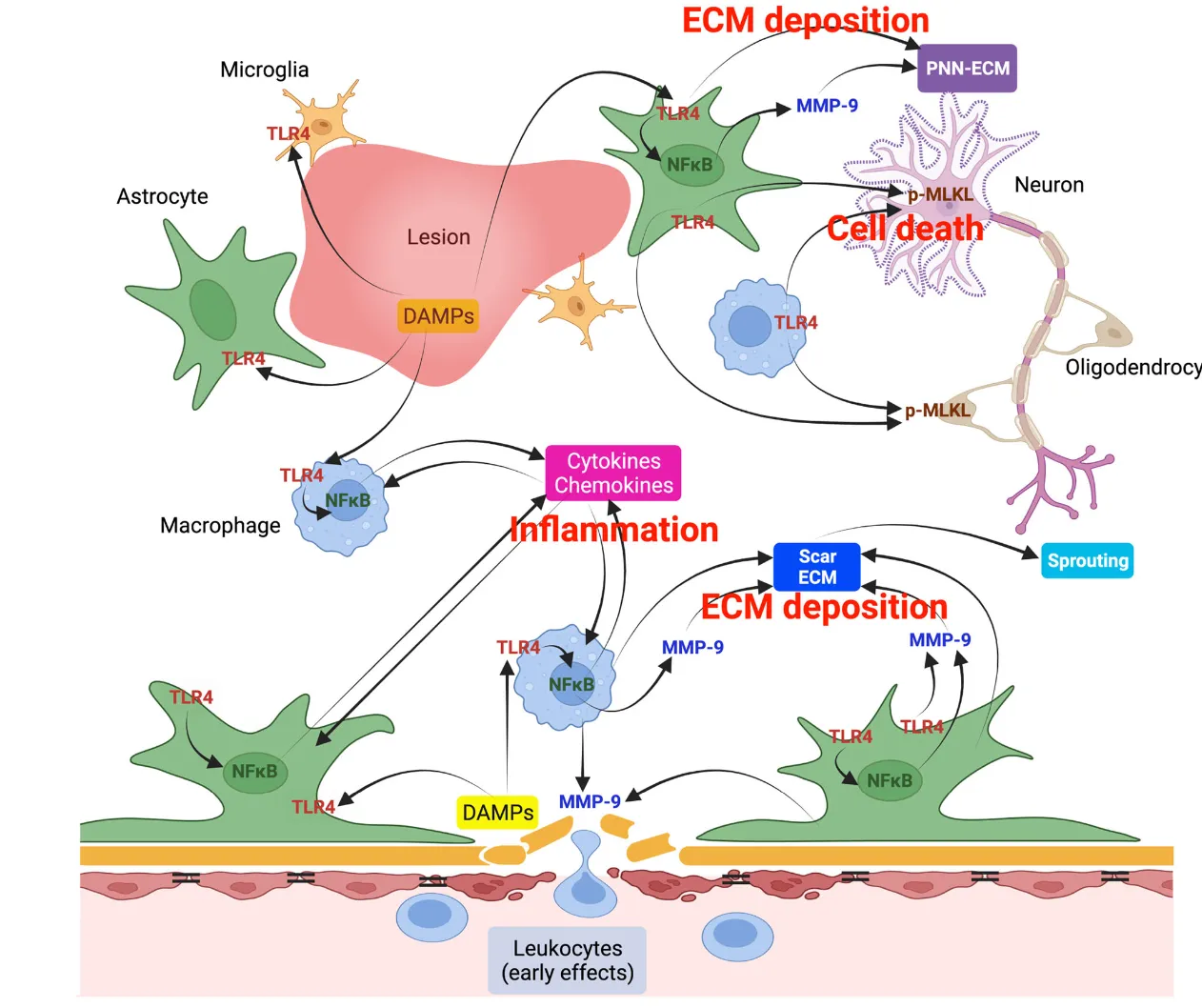
The reason RIPK3 and MLKL are being investigated is that they are involved in a cell death pathway called necroptosis. Necroptosis is a form of inflammatory cell death and may play an important role in the inflammatory response and cell death that follow spinal cord injury.
Specifically, phosphorylation of RIPK3 activates MLKL, which translocates to the cell membrane and causes its disruption. This not only kills the cell but also releases damage-associated molecular patterns (DAMPs), which sustain a further TLR4-mediated inflammatory response.
This study shows that, by evaluating the effect of TLR4 deficiency on this necroptosis pathway after spinal cord injury, it may be possible to reduce chronic inflammation and secondary damage and to promote neuroprotection and functional recovery.
The link between RIPK3/MLKL and TLR4
RIPK3 and MLKL are linked to TLR4 because TLR4 may regulate the necroptosis pathway through these molecules.
TLR4 is a key receptor that triggers the inflammatory response and is involved in chronic inflammation after spinal cord injury. RIPK3 and MLKL are central molecules in the necroptosis pathway induced by TLR4 activation. In this pathway, TLR4 signaling promotes phosphorylation of RIPK3, which in turn triggers activation of MLKL. Activated MLKL translocates to the cell membrane and induces cell death through membrane disruption.
This study shows that TLR4 deficiency suppresses the activity of RIPK3 and MLKL, thereby reducing necroptosis and suppressing chronic inflammation. As a result, loss of neurons and myelin is reduced, and recovery of motor function is promoted, as confirmed in the study.
Why this study focused on TLR4
This study focused on TLR4 because TLR4 plays an important role in the inflammatory response after spinal cord injury. Specifically, TLR4 drives the production of inflammatory cytokines and changes in the extracellular matrix (ECM), which can worsen secondary tissue damage and functional impairment.
The underlying problem
After spinal cord injury, local tissue damage occurs during the initial acute phase, followed by a secondary inflammatory response that persists over the long term. This chronic inflammatory response inhibits the regeneration of nervous tissue and becomes a factor that impedes recovery.
The role of TLR4
As a pattern recognition receptor that triggers inflammation, TLR4 recognizes foreign pathogens and endogenous damage-associated molecules (DAMPs). After spinal cord injury, TLR4 is thought to become excessively activated, inducing chronic inflammation and cell death.
Aim of the study
The aim of this study is to elucidate how TLR4 is involved in the inflammatory response and cell death in chronic-phase spinal cord injury, and how this affects secondary damage and functional recovery. It is hoped that this will lead to the development of new therapies targeting TLR4.
TLR4-expressing cells in the chronic phase of spinal cord injury
In the chronic phase of spinal cord injury (8 weeks later), the cells expressing TLR4 are, in order of abundance, as follows.
- Astrocytes (GFAP-positive cells)
TLR4 is particularly strongly expressed in astrocytes during the chronic phase, and its expression level increases further over time. - Macrophages/microglia (CD11b-positive cells)
TLR4 expression is also confirmed in macrophages and microglia, but in the chronic phase this expression is not as strong as in astrocytes and is reduced compared with day 7. - Neurons (NeuN-positive cells)
Little TLR4 expression is seen in neurons.
Thus, in the chronic phase after spinal cord injury, TLR4 is mainly highly expressed in astrocytes, which are thought to be involved in inflammation and tissue remodeling.
What are CC1-positive cells?
CC1-positive cells refer to oligodendrocytes. Oligodendrocytes are cells that form the myelin sheath in the central nervous system (CNS); they provide insulation to neuronal axons and serve to improve the conduction speed of nerve signals.
CC1 is widely used as an oligodendrocyte-specific marker and is employed for identifying oligodendrocytes and quantifying their numbers. In studies after spinal cord injury, because the number and function of oligodendrocytes are important for nerve regeneration and recovery, analysis of CC1-positive cells is performed.
Off-target effects of TLR4-KO
The possible off-target effects when using TLR4-KO mice are as follows.
- Cross-reactivity with other TLR family members
TLR4 is a receptor belonging to the TLR family, and its signaling pathways may overlap with those of other TLRs (such as TLR2 and TLR3). When TLR4 is deficient, other TLRs may be compensatorily activated, which could affect the observed effects. - Overall suppression of the inflammatory response
Because TLR4 is a major regulator of the inflammatory response, its deficiency may suppress the overall immune response. As a result, inflammation and immune responses that do not directly depend on TLR4 may also be affected, potentially producing anomalous results. - Effects on metabolism and other cellular functions
Because TLR4 is involved not only in the inflammatory response but also in cellular metabolism and survival signaling, TLR4 deficiency may affect metabolic pathways and basic cellular functions. This may give rise to off-target effects in contexts other than spinal cord injury. - Influence of genetic background
Because TLR4-KO mice carry a specific genetic background, interactions with other genes may produce unexpected biological effects. In particular, if backcrossing is incomplete, the influence of background genes may become pronounced.
These off-target effects suggest that the observations attributable to TLR4 deficiency may not necessarily be due to TLR4 itself
. Therefore, interpretation of the results requires caution, and it is desirable to verify the results using other methods (e.g., conditional knockout, the use of TLR4 inhibitors).
Site of mRNA extraction and RNA sequencing
In this study, mRNA was extracted from the spinal cord injury site and RNA sequencing (RNA-seq) was performed. Specifically, about 5mm of spinal cord tissue including the center of the injury (the epicenter) was collected on day 7 and at 8 weeks after injury, and mRNA was extracted from that tissue. Using this approach, changes in gene expression at the injury site were analyzed in detail to investigate how TLR4 deficiency affects the inflammatory response and extracellular matrix (ECM)–related genes after spinal cord injury.
Whether scRNA-seq was used
In this study, scRNA-seq (single-cell RNA sequencing) was not performed. What was used is RNA-seq, which targets the mRNA of whole tissue extracted from the spinal cord injury site.
RNA-seq is a method for examining the gene expression of the whole tissue, and it does not analyze differences in gene expression at the level of individual cells as scRNA-seq does. Therefore, this study does not analyze detailed gene expression patterns for each individual cell type, but rather captures the overall changes in gene expression after spinal cord injury.
Explanation of Fig 4
Figure 5 is a figure comparing NFκB signaling and the expression of inflammatory cytokines/chemokines after spinal cord injury in TLR4-deficient (TLR4 KO) mice and wild-type (WT) mice. This figure mainly shows the following points.
Activation of NFκB signaling
- Fig 5A, B:
Nuclear translocation of NFκB-P65 is compared between TLR4 KO and WT mice. NFκB-P65 is the central transcription factor of the NFκB pathway and regulates the inflammatory response. In TLR4 KO mice, nuclear translocation of NFκB-P65 after spinal cord injury is suppressed, indicating that NFκB signaling activation is lower than in WT mice. - Fig 5C, D:
The process by which NFκB is activated through phosphorylation of IκBα (p-IκBα) and its degradation is shown. In TLR4 KO mice, the p-IκBα/IκBα ratio is lower than in WT mice, confirming that activation of NFκB is suppressed.
Expression of inflammatory cytokines and chemokines
- Fig 5E-H:
The mRNA expression levels of inflammatory cytokines and chemokines such as IL-1β, TNFα, IL-6, and CCL2 are shown. In WT mice, the expression of these inflammatory molecules increases after spinal cord injury, but in TLR4 KO mice this expression is significantly suppressed. This suggests that TLR4 contributes to the strengthening of the inflammatory response.
Summary
Figure 5 shows that TLR4 plays an important role in the inflammatory response after spinal cord injury, revealing that TLR4 deficiency leads to suppression of NFκB signaling and the accompanying reduction in the expression of inflammatory cytokines and chemokines. This suggests that TLR4-targeted therapy may be effective in controlling chronic inflammation after spinal cord injury.
What is t-SNE flow cytometry analysis?
t-SNE (t-distributed Stochastic Neighbor Embedding) flow cytometry analysis is a method for visualizing multidimensional data in a two- or three-dimensional space. In particular, it is widely used in the analysis of flow cytometry data to find patterns and subsets within complex cell populations.
About the specific method
- Flow cytometry:
Flow cytometry is a technique that measures the physical and chemical properties of cells, capable of simultaneously measuring numerous parameters such as cell size, shape, and the expression of surface and internal molecules. - The principle of t-SNE:
t-SNE is a nonlinear method for mapping high-dimensional data into a low-dimensional space. While preserving the similarities in the original data space, it places data points in a two- or three-dimensional plot. With this method, similar cells gather close together, while different cell populations are placed far apart.
Applications of t-SNE flow cytometry analysis
By applying this method to flow cytometry, the complex relationships among cell populations can be visually analyzed based on multiple parameters. Specifically, based on the expression of cell-surface markers and internal markers, it is possible to identify subsets of different immune cells and to evaluate their dynamics in detail.
The role of t-SNE flow cytometry analysis in this study
In this study, t-SNE–based flow cytometry analysis was used to visualize the diversity and dynamics of immune cells in TLR4-deficient mice and wild-type mice after spinal cord injury, analyzing differences and changes by cell population in detail. This makes it possible to gain a deeper understanding of the effect of TLR4 on the immune response after injury.
Antibodies used
In the flow cytometry (FCM) analysis used in this study, the following antibodies were used to identify and characterize immune cells. These antibodies are designed to detect specific cell-surface markers and internal markers.
List of antibodies used
- CD45 (BUV395, 1:300, BD Biosciences):
Antibody for identifying all leukocytes (immune cells). - CD11b (BV421; 1:300; BD Biosciences):
Expressed on the surface of myeloid cells such as macrophages and microglia. - Ly6G (BUV737; 1:400; BD Biosciences):
A marker for identifying mainly neutrophils. - F4/80 (APC; 1:250; BD Biosciences):
A marker specific to macrophages. - Ly6C (BV711; 1:300; BD Biosciences):
Identifies monocytes and a subset of T cells. - CD11c (BV650; 1:250; BD Biosciences):
A marker for dendritic cells (DCs). - MHC-II (FITC; 1:300; BD Biosciences):
Expressed on the surface of antigen-presenting cells (macrophages, dendritic cells, B cells, etc.). - iNOS (PerCP; 1:250; BD Biosciences):
The inducible form of nitric oxide synthase (expressed in inflammatory macrophages and other
cells).
- ArgI (PE; 1:200; BD Biosciences):
Arginase I, a marker of M2-type macrophages. - CD206 (PE-Cy7; 1:350; BD Biosciences):
Expressed on the surface of M2-type macrophages and dendritic cells. - CD19 (BV605; 1:250; BD Biosciences):
A marker for B cells. - CD3 (AF700; 1:200; BD Biosciences):
A surface marker of all T cells. - CD4 (APC-Cy7; 1:200; BD Biosciences):
A marker for helper T cells. - CD8 (PE-Dazzle594; 1:250; BD Biosciences):
A marker for killer T cells. - Live/dead UV (1:1,000; BioLegend):
Staining to distinguish dead cells from live cells.
The purpose of using these antibodies
These antibodies are used to identify subsets of different immune cells and to analyze their dynamics and characteristics in detail. Specifically, these antibodies are used to clarify the differences in the immune response after spinal cord injury between TLR4-deficient mice and wild-type mice.
Can they be measured simultaneously?
Normally, in flow cytometry (FCM) it is possible to use multiple antibodies simultaneously to measure various cell-surface markers and internal markers at once. This process is called “multicolor flow cytometry.” However, the number of antibodies that can be measured simultaneously depends on the performance of the flow cytometer used.
Specifically:
- Number of lasers and detectors on the flow cytometer:
A typical flow cytometer is equipped with multiple lasers and detectors, each able to excite and detect fluorescence at different wavelengths. Each antibody is labeled with a different fluorescent dye (fluorophore), and each is excited by a specific laser and emits at a different wavelength. - Spectral overlap:
As the number of antibodies used increases, spectral overlap between fluorescent dyes can become a problem. To mitigate this problem, fluorescence compensation is required. - Panel design:
With effective panel design, multiple antibodies can be used simultaneously. The antibody set used in this study can be measured simultaneously on many flow cytometers.
Summary
The number of antibodies used in this study can be measured simultaneously using a multicolor flow cytometer. However, the actual number that can be measured simultaneously depends on the specifications of the flow cytometer. The latest flow cytometers can also measure more than 20 antibodies simultaneously, but appropriate panel design and compensation to match this are required.
Molecules examined as ECM
In this study, the expression of extracellular matrix (ECM)–related molecules after spinal cord injury was analyzed in TLR4-deficient mice and wild-type mice. The specific ECM-related molecules examined are as follows.
- Versican:
A large proteoglycan in the ECM, involved in cell adhesion, migration, and intercellular signaling. - Lumican:
A small lipopolysaccharide protein, related to collagen formation and cell migration. - Phosphacan (PTPRZ):
A proteoglycan abundant in nervous tissue, involved in signaling in the extracellular region. - Decorin:
A small proteoglycan that, through interaction with collagen fibers, regulates the structure and function of tissue. - Collagen 1A1 (COL1A1):
A major component of collagen, providing tissue strength and flexibility. - Matrix Metalloproteinase-9 (MMP9):
Involved in the degradation of the ECM and plays an important role in tissue remodeling after injury.
Aim of the study
By examining these ECM molecules, the study evaluates how TLR4 deficiency affects ECM remodeling and tissue regeneration after injury. In particular, changes in the ECM are deeply involved in nerve regeneration and the progression of secondary damage after spinal cord injury, making this important for understanding how TLR4 affects these processes.
Results for Collagen 1
The results for Collagen 1A1 (COL1A1) in this study examine the difference in expression after spinal cord injury between TLR4-deficient mice (TLR4 KO mice) and wild-type mice.
Overview of the results
In TLR4 KO mice, expression of Collagen 1A1 at 8 weeks after spinal cord injury was shown to be lower than in wild-type mice. This result suggests that TLR4 contributes to ECM remodeling and collagen accumulation. In particular, TLR4 deficiency may suppress the abnormal accumulation of ECM molecules, potentially reducing inflammatory responses and scar formation.
Specific data
In Western blot and mRNA analyses, the expression level of COL1A1 in TLR4 KO mice was shown to be significantly lower than in wild-type mice. This reveals that TLR4 plays an important role in collagen accumulation after spinal cord injury in the chronic phase.
Significance
The reduced expression of COL1A1 suggests that TLR4 deficiency suppresses excessive scar formation of tissue and may promote nerve regeneration and functional recovery. This indicates that TLR4-targeted therapies are promising as a treatment strategy in the chronic phase after spinal cord injury.