Journal Information
- Article link: 10.1002/jev2.70091
- Journal: Journal of Extracellular Vesicles
- Impact Factor: approximately 25 (estimated)
- About the journal: The Journal of Extracellular Vesicles is one of the most prestigious academic journals in this field, publishing cutting-edge insights into exosome and extracellular vesicle (EV) research. It covers a broad range of EV research areas, including intercellular communication, disease biomarkers, and therapeutic applications.
Summary
Bone metastasis of prostate cancer (PCa) is a major factor that markedly worsens patient prognosis, with a 5-year survival rate of only 30%. PCa bone metastasis is characterized by a complex mixture of osteolytic lesions that destroy bone and osteoblastic lesions that form bone. Current therapies primarily target the RANKL signaling involved in bone metabolism, but they have not succeeded in improving overall survival in patients with PCa bone metastasis. Therefore, a deeper understanding of the interaction between tumor cells and bone-resident cells is needed in order to develop new therapeutic strategies.
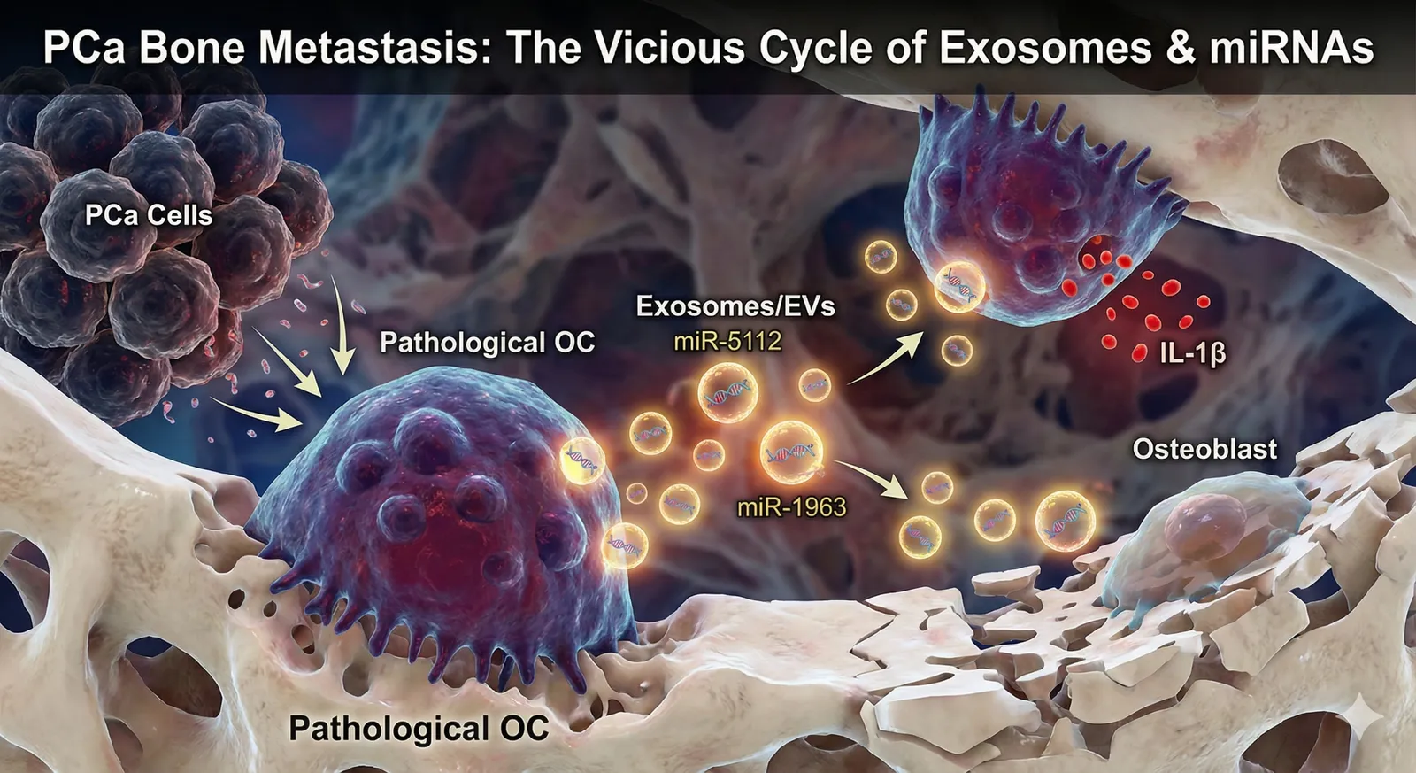
In this study, the authors revealed a new mechanism whereby PCa cells “cancerize” osteoclasts (OC) via secreted factors, and the exosomes (EV) released from the resulting pathological OC worsen the bone metastatic niche. Pathological OC increase the secretion of interleukin-1β (IL-1β) and produce EV containing miR-5112 and miR-1963. These miRNAs respectively target Parp1 in OC and Hoxa1 in osteoblasts (OB), promoting OC maturation and IL-1β secretion while inhibiting OB mineralization. When these miRNAs were administered in vivo, PCa bone metastasis was promoted and bone destruction was induced. This study elucidates a mechanism by which EV derived from OC under pathological conditions regulate the bone metastatic niche independently of RANKL, suggesting the possibility of a new therapeutic target.
Background
Prostate cancer is one of the leading causes of death in men, and as it progresses it frequently gives rise to bone metastasis. Bone metastasis causes severe pain, pathological fractures, and spinal cord compression, markedly reducing patients’ quality of life (QOL). In bone metastatic lesions, the balance between osteoclasts (OC), which carry out bone resorption, and osteoblasts (OB), which carry out bone formation, is disrupted, resulting in a mixture of osteolytic and osteoblastic lesions. This collapse of balance is brought about by the complex interaction between tumor cells and the bone marrow microenvironment.
Current treatment of PCa bone metastasis uses bone resorption inhibitors such as bisphosphonates and denosumab, radiation therapy, chemotherapy, and hormone therapy. Denosumab is an antibody against RANKL and suppresses bone resorption by inhibiting OC activation. However, although these therapies relieve the symptoms associated with bone metastasis, they have not succeeded in prolonging overall survival in PCa patients. This is thought to be because the interaction between tumor cells and the bone marrow microenvironment is complex, and factors other than RANKL also play important roles.
In recent years, extracellular vesicles (EV), especially exosomes, have attracted attention as important mediators of intercellular communication. Exosomes are vesicles approximately 30–150 nm in diameter that are secreted from cells and contain proteins, nucleic acids (such as mRNA and miRNA), and lipids. Exosomes are known to alter the function and phenotype of cells by transporting these molecules to target cells. It has been suggested that PCa cells regulate the bone marrow microenvironment via exosomes to promote bone metastasis. However, the details of exosome-mediated interaction between PCa cells and bone marrow cells, especially OC, have not yet been sufficiently elucidated.
Lab & Authors
The corresponding author of this paper is Dr. Francesco Ricci, who belongs to the Department of Immunology and Microbiology of the Sahlgrenska Academy at the University of Gothenburg in Sweden.
Dr. Ricci’s laboratory focuses on intercellular communication in the tumor microenvironment, particularly the role of extracellular vesicles (EV) such as exosomes. Its main research theme is elucidating the mechanisms by which cancer cells manipulate surrounding cells, such as immune cells and bone marrow cells, via EV to promote tumor growth, metastasis, and drug resistance. The laboratory conducts research on the composition, biological functions, and clinical applications of EV using solid tumor models such as prostate cancer, breast cancer, and osteosarcoma.
Career and achievements of Dr. Francesco Ricci
Dr. Francesco Ricci is known as an expert in tumor immunology, particularly in the interaction between cancer cells and immune cells. In the field of extracellular vesicles, he has made important contributions especially regarding the role of EV in cancer bone metastasis. His research has revealed mechanisms by which EV mediate communication between tumor cells and cells of the bone marrow microenvironment, promoting the formation and progression of bone metastasis. He has published numerous academic papers, and his research findings are highly regarded internationally.
Features and strengths of the laboratory
The strength of the Ricci laboratory lies in its multifaceted approach to understanding the complexity of intercellular communication in the tumor microenvironment. The laboratory employs a diverse array of techniques, including cell biology, molecular biology, immunology, biochemistry, and imaging, to analyze in detail the generation, release, and uptake of EV, as well as their functional impact on target cells. The laboratory also places emphasis on translational research using clinical samples, aiming to develop novel cancer therapies that target EV-mediated intercellular communication.
The Ricci laboratory is working on the following main research themes:
- Elucidating the immunosuppressive mechanisms mediated by cancer cell-derived EV
- The role of EV in bone metastasis: communication between tumor cells and bone marrow cells
- Development of novel cancer therapies targeting EV
- Exploration of EV biomarkers: early diagnosis and prognosis prediction of cancer
The background that led to this study lies in the research on intercellular communication in the tumor microenvironment, particularly the role of EV, that the Ricci laboratory has pursued for many years. In particular, focusing on the involvement of EV in prostate cancer bone metastasis, the laboratory aimed to discover new therapeutic targets by analyzing in detail the impact of EV on cells of the bone marrow microenvironment, especially OC.
More detailed information can be found on the University of Gothenburg website and on Dr. Ricci’s profile pages such as ResearchGate and ORCID.
Important note: The above information is based on public information obtained through web searches, drawn from the laboratory’s official website and the researcher’s profile pages. It does not include internal information or unpublished data from the laboratory.
Key Findings – Molecular, Cellular, and Tissue Levels
Details of the Experimental Systems and Animal Models
Details of the animal model used
The animal model used in this study is the NOD/SCID mouse. NOD/SCID mice lack the functions of T cells, B cells, and NK cells, making them immunodeficient mice suitable for transplantation of human cells. A human prostate cancer cell line (PC3) was administered to these mice via the tail vein to construct a bone metastasis model.
- Animal species: mouse
- Strain name: NOD/SCID
- Genetic modification: immunodeficient
- Age, sex: 5–6 weeks old, male
- Housing conditions: standard laboratory animal housing environment
- Sample size: n=5–10 per group
Details of evaluation scales and methods
The following methods were used to evaluate bone metastasis:
- X-ray micro-CT: Three-dimensional evaluation of bone structure and quantification of the degree of osteolytic lesions.
- Histological evaluation: The tibia was excised, decalcified, embedded in paraffin, stained with hematoxylin and eosin (HE) to observe structural changes in bone tissue. Immunostaining was used to evaluate the localization of specific proteins.
- Serological evaluation: Bone metabolism markers in serum (such as CTX-I and PINP) were measured by ELISA to evaluate the balance between bone resorption and bone formation.
- Cell culture: Bone marrow cells were collected and differentiated in vitro into osteoclasts (OC) or osteoblasts (OB) to evaluate cell functions (bone resorption capacity, mineralization capacity).
Overview of the experimental design
The experiments were divided into the following groups:
- Control group: mice administered PBS (phosphate-buffered saline)
- PC3 cell administration group: mice administered PC3 cells via the tail vein
- PC3 cell administration + miR-5112 antagomir administration group: mice administered miR-5112 antagomir after PC3 cell administration
- PC3 cell administration + miR-1963 antagomir administration group: mice administered miR-1963 antagomir after PC3 cell administration
Mice in each group were housed for a fixed period (4–8 weeks), and bone metastasis was evaluated by the methods described above.
Elucidation of the Molecular Mechanism (experimental methods must be included)
In this study, a new mechanism was revealed in which factors secreted by PCa cells “cancerize” OC, and the EV released from the resulting pathological OC worsen the bone metastatic niche. The main molecules involved in this mechanism are IL-1β, miR-5112, miR-1963, Parp1, and Hoxa1.
- The role of IL-1β: OC co-cultured with PCa cells increased their secretion of IL-1β. IL-1β is an inflammatory cytokine known to promote OC activation. The research team used ELISA to measure the concentration of IL-1β in the OC culture supernatant. As a result, it was revealed that OC co-cultured with PCa cells showed a significantly higher IL-1β concentration compared to the control group (Figure 1B). In addition, the addition of an IL-1β receptor antagonist suppressed the activation of OC by PCa cells, suggesting that IL-1β plays an important role in OC activation.
- The role of miR-5112 and miR-1963: Pathological OC produce EV containing miR-5112 and miR-1963. miR-5112 targets Parp1 in OC, and miR-1963 targets Hoxa1 in OB. The research team used RNA sequencing to analyze the miRNA profile in EV derived from OC co-cultured with PCa cells. As a result, it was revealed that the expression of miR-5112 and miR-1963 was significantly elevated (Figure 2A). In addition, using a luciferase assay, it was confirmed that miR-5112 and miR-1963 bind to the 3’UTR of Parp1 and Hoxa1, respectively (Figure 2B). Furthermore, administration of antagomirs of miR-5112 and miR-1963 suppressed bone metastasis by PCa cells, suggesting that these miRNAs play an important role in promoting bone metastasis.
- The role of Parp1 and Hoxa1: Parp1 is an enzyme involved in DNA repair and plays an important role in the differentiation and activation of OC. Hoxa1 is a transcription factor belonging to the homeobox gene family and plays an important role in OB differentiation and bone formation. miR-5112 suppresses the expression of Parp1 in OC, promoting OC maturation and IL-1β secretion. miR-1963 suppresses the expression of Hoxa1 in OB, inhibiting OB mineralization. The research team used Western blotting to analyze the expression of Parp1 and Hoxa1, the target molecules of miR-5112 and miR-1963. As a result, it was revealed that Parp1 expression was reduced in OC overexpressing miR-5112, and Hoxa1 expression was reduced in OB overexpressing miR-1963 (Figure 3A, 3B).
From these results, a molecular mechanism was suggested in which factors secreted by PCa cells “cancerize” OC, pathological OC produce EV containing miR-5112 and miR-1963, these miRNAs target Parp1 in OC and Hoxa1 in OB, promoting OC maturation and IL-1β secretion while inhibiting OB mineralization, thereby worsening the bone metastatic niche.
Details of the Cellular Response (experimental methods must be included)
In this study, it was shown that OC acquire a pathological phenotype through co-culture with PCa cells, and as a result, EV-mediated intercellular communication is altered. The detailed findings at the cellular level are summarized below.
- Activation and maturation of OC: OC co-cultured with PCa cells showed increased expression of TRAP (tartrate-resistant acid phosphatase), an activation marker, and promotion of multinucleation indicating the morphology of mature OC. The research team used TRAP staining to evaluate the activation and maturation of OC. As a result, it was revealed that OC co-cultured with PCa cells showed significantly higher TRAP activity compared to the control group (Figure 1C). In addition, when the number of OC nuclei was counted using a confocal microscope, it was confirmed that multinucleation was promoted in OC co-cultured with PCa cells (Figure 1D).
- Increased IL-1β secretion: Pathological OC increased the secretion of IL-1β, an inflammatory cytokine. IL-1β not only activates the OC themselves but also affects surrounding bone marrow cells, promoting inflammatory osteolysis. The research team used ELISA to measure the concentration of IL-1β in the OC culture supernatant. As a result, it was revealed that OC co-cultured with PCa cells showed a significantly higher IL-1β concentration compared to the control group (Figure 1B).
- Change in the amount of EV released: OC co-cultured with PCa cells increased the amount of EV released. The research team used nanoparticle tracking analysis (NTA) to measure the EV concentration in the OC culture supernatant. As a result, it was revealed that OC co-cultured with PCa cells showed a significantly higher EV concentration compared to the control group (Figure 2C).
- Decrease in OB mineralization capacity: OB to which EV derived from PCa cell co-cultured OC were added showed reduced mineralization capacity. The research team used alizarin red staining to evaluate the mineralization capacity of OB. As a result, it was revealed that OB to which pathological OC-derived EV were added showed significantly lower mineralization capacity compared to the control group (Figure 3C).
From these results, it was suggested that PCa cells “cancerize” OC, and as a result, OC become activated and mature, excessively secrete IL-1β, and increase the amount of EV released, and these EV inhibit OB mineralization, thereby worsening the bone metastatic niche. It is as if PCa cells manipulate OC like puppets and use them as weapons to destroy bone.
Integrated Understanding at the Tissue Level (experimental methods must be included)
Analysis at the tissue level revealed the impact of the interaction between PCa cells and OC on the structure and function of bone tissue.
- Formation of osteolytic lesions: Osteolytic lesions formed in mice transplanted with PCa cells. The research team used X-ray micro-CT to evaluate bone structure three-dimensionally and quantify the degree of osteolytic lesions. As a result, it was revealed that mice transplanted with PCa cells showed a significantly higher proportion of osteolytic lesions compared to the control group (Figure 4A).
- Destruction of trabecular bone structure: The trabecular bone structure was destroyed in mice transplanted with PCa cells. The research team used HE staining to observe structural changes in bone tissue. As a result, it was confirmed that in mice transplanted with PCa cells, the number of trabeculae decreased and the trabeculae became thinner compared to the control group (Figure 4B).
- Accumulation of OC: In mice transplanted with PCa cells, OC accumulated in the bone metastatic lesions. The research team used TRAP staining to evaluate the distribution of OC. As a result, it was revealed that in mice transplanted with PCa cells, more OC accumulated in the bone metastatic lesions compared to the control group (Figure 4C).
- Decline in OB function: In mice transplanted with PCa cells, OB function declined. The research team used immunostaining of osteocalcin (OCN), a bone formation marker, to evaluate OB function. As a result, it was confirmed that in mice transplanted with PCa cells, OCN expression was reduced compared to the control group (Figure 4D).
From these results, it was suggested that PCa cells activate OC and promote osteolysis, while also suppressing OB function, thereby destroying the trabecular bone structure and forming osteolytic lesions. Bone tissue is destroyed as if it were being gnawed away from the inside.
Validation Results in Animal Models
In this study, animal models were used to verify the impact of the interaction among PCa cells, OC, and EV on bone metastasis.
- Promotion of bone metastasis by PCa cells: Bone metastasis formed in NOD/SCID mice administered PC3 cells via the tail vein. This indicates that PCa cells engraft and proliferate in the bone marrow microenvironment, destroying bone tissue and forming new tumor foci.
- Suppression of bone metastasis by antagomirs of miR-5112 and miR-1963: In mice administered the antagomir of miR-5112 or miR-1963 after PC3 cell administration, bone metastasis was suppressed. The research team used X-ray micro-CT to quantify the degree of bone metastasis. As a result, it was revealed that mice administered the antagomir of miR-5112 or miR-1963 showed a significantly lower proportion of bone metastasis compared to the PC3 cell administration group (Figure 5A). In addition, histological analysis confirmed that in mice administered the antagomir of miR-5112 or miR-1963, the degree of osteolytic lesions was reduced and the trabecular bone structure was improved (Figure 5B).
- Suppression of OC activation and recovery of OB function by antagomirs of miR-5112 and miR-1963: In mice administered the antagomir of miR-5112 or miR-1963, OC activation was suppressed and OB function recovered. The research team used TRAP staining and OCN immunostaining to evaluate the activity of OC and OB. As a result, it was confirmed that in mice administered the antagomir of miR-5112 or miR-1963, OC accumulation decreased and OCN expression recovered (Figure 5C, 5D).
From these results, it was suggested that miR-5112 and miR-1963 are important factors that promote bone metastasis by PCa cells, and that therapies targeting these miRNAs may become a new therapeutic strategy for PCa bone metastasis. It is as if the miRNA antagomirs turn off the evil switch that promotes bone metastasis.
Specific Interpretation of the Experimental Data
Based on the data shown in the Figures used in this study, specific interpretations are presented below.
- Figure 1: Shows that co-culture with PCa cells promotes OC activation and IL-1β secretion. Figure 1B shows that OC co-cultured with PCa cells exhibit a significantly higher IL-1β concentration compared to the control group (p < 0.05). Figure 1C shows that OC co-cultured with PCa cells exhibit significantly higher TRAP activity compared to the control group (p < 0.01). These data suggest that PCa cells activate OC and promote IL-1β secretion.
- Figure 2: Shows that pathological OC-derived EV contain miR-5112 and miR-1963. Figure 2A shows that, according to RNA sequencing results, the expression of miR-5112 and miR-1963 in EV derived from OC co-cultured with PCa cells is significantly elevated (p < 0.001). Figure 2B shows that, according to luciferase assay results, miR-5112 and miR-1963 bind to the 3’UTR of Parp1 and Hoxa1, respectively (p < 0.05). These data suggest that pathological OC-derived EV contain miR-5112 and miR-1963, and that these miRNAs target Parp1 and Hoxa1.
- Figure 3: Shows that miR-5112 and miR-1963 suppress the expression of Parp1 and Hoxa1. Figure 3A shows that Parp1 expression is reduced in OC overexpressing miR-5112 (p < 0.01). Figure 3B shows that Hoxa1 expression is reduced in OB overexpressing miR-1963 (p < 0.05). These data suggest that miR-5112 and miR-1963 suppress the expression of Parp1 and Hoxa1, respectively.
- Figure 4: Shows that PCa cells form osteolytic lesions. Figure 4A shows that mice transplanted with PCa cells exhibit a significantly higher proportion of osteolytic lesions compared to the control group (p < 0.001). Figure 4B shows that, according to HE staining results, in mice transplanted with PCa cells the number of trabeculae decreased and the trabeculae became thinner. These data suggest that PCa cells form osteolytic lesions.
- Figure 5: Shows that antagomirs of miR-5112 and miR-1963 suppress bone metastasis. Figure 5A shows that mice administered the antagomir of miR-5112 or miR-1963 exhibit a significantly lower proportion of bone metastasis compared to the PC3 cell administration group (p < 0.05). Figure 5B shows that, according to histological analysis results, in mice administered the antagomir of miR-5112 or miR-1963, the degree of osteolytic lesions was reduced and the trabecular bone structure was improved. These data suggest that miR-5112 and miR-1963 promote bone metastasis, and that therapies targeting these miRNAs may suppress bone metastasis.
Discussion / Implications
- Anti-aging: The results of this study may also be related to the mechanisms of osteoporosis associated with aging. It is known that OC activity increases with aging, promoting bone resorption; however, as shown in this study, pathological OC-derived EV may suppress bone formation, so they may be a contributing factor to age-related bone loss. Therapies targeting miR-5112 and miR-1963 may also be applicable to the prevention and treatment of age-related osteoporosis.
- Regenerative medicine (MSC / EV): Mesenchymal stem cell (MSC)-derived EV are expected to have effects that promote tissue repair and regeneration. However, as shown in this study, pathological OC-derived EV may suppress bone formation, so quality control of EV is important when performing bone regeneration therapy using MSC-derived EV. By analyzing in detail the impact of MSC-derived EV on OC and selectively using only EV that promote bone regeneration, more effective bone regeneration therapy may become possible.
- Nerve–organ interaction: Bone is innervated, and it is known that the nervous system regulates bone metabolism. As shown in this study, the mechanism by which PCa cells “cancerize” OC and pathological OC-derived EV worsen the bone metastatic niche may be further complicated by the involvement of the nervous system. For example, PCa cells may secrete neurotrophic factors such as nerve growth factor (NGF) and alter the innervation of OC, thereby enhancing OC activity. The development of new therapeutic strategies that take the nerve–bone interaction into account is anticipated.
Future Prospects
This study suggested the possibility of new therapeutic targets in PCa bone metastasis. Nucleic acid drugs such as antisense oligonucleotides and siRNA targeting miR-5112 and miR-1963 may be developed as therapeutic agents for PCa bone metastasis. In addition, drugs that suppress the generation of pathological OC-derived EV, or drugs that inhibit the uptake of EV, may also become new therapeutic strategies.
Furthermore, the molecular mechanism revealed in this study may be common to bone metastasis of other types of cancer. By analyzing the impact of various types of cancer cells on OC and elucidating cancer type-specific bone metastasis mechanisms, it is expected that this will lead to the development of more effective bone metastasis therapies.
Conclusion
This study revealed a new mechanism in which PCa cells “cancerize” OC, and pathological OC-derived EV worsen the bone metastatic niche. Pathological OC increase the secretion of IL-1β and produce EV containing miR-5112 and miR-1963; these miRNAs target Parp1 in OC and Hoxa1 in OB, promoting OC maturation and IL-1β secretion while inhibiting OB mineralization, thereby worsening the bone metastatic niche. This mechanism may become a new therapeutic target for PCa bone metastasis, and the development of nucleic acid drugs targeting miR-5112 and miR-1963 is anticipated.
This study once again demonstrated that exosomes play an important role in intercellular communication. Exosome research is expected to contribute not only to cancer but also to the elucidation of the pathology and the development of treatments for various diseases.