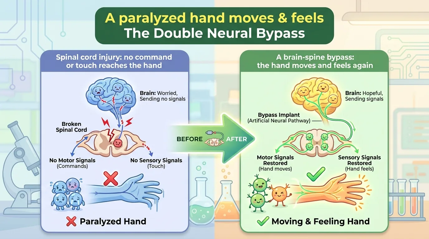
You think, “I’ll move my hand.” That thought becomes an electrical signal in the brain’s motor cortex, travels down the thick neural cable of the spinal cord, and reaches the muscles of the arm and fingers. This sequence, which we all perform without a second thought, works only because it is connected by a single road called the spinal cord. Conversely, when a fingertip touches something, that sensation travels up from the peripheral nerves through the spinal cord, reaches the brain’s sensory cortex, and becomes the felt reality of “I touched it.” The outbound motor command and the inbound sensory information. The brain and the hand are bound together by this two-way exchange.
Spinal cord injury (SCI) is an injury that severs this road. Especially when it occurs high in the neck and the nerves are completely cut—a state of “complete paralysis”—no matter how hard the brain wills the hand to “move,” the command never reaches it, and whatever the hand touches, that sensation never rises to the brain. Movement and sensation are lost together. Restoring hand function is always ranked first among the recoveries that the people affected most urgently want.
And by conventional medical wisdom, complete-paralysis spinal cord injury has been held to be beyond recovery. The paper introduced today challenges that wisdom head-on. A man paralyzed almost completely from the neck down came to move his own hand by thought alone, to grip an eggshell without breaking it, to feed himself, and—to regain touch in a wrist whose connection was supposedly severed, keeping it even after the device was switched off. This is the story of a “double neural bypass” that bridges the brain to the spinal cord and cerebral cortex at the same time.
Journal Information
- Paper title: A neuroprosthesis for restoring hand movement and sensation in a person with complete tetraplegia
- Authors: Santosh Chandrasekaran and Sarah K. Wandelt (the two lead authors, equal contribution), and others: Aniket Jangam, Zeev Elias, Erona Ibroci, Christina Maffei, Isabelle A. Rosenthal, Richard Ramdeo, Joo-won Kim, Junqian Xu, Matthew F. Glasser, Allison Neuwirth, Todd A. Goldstein, Nathan E. Crone, Matthew F. Fifer, Gelana Tostaeva, Stephan Bickel, Douglas Griffin, Michael Funaro, Nicholas G. Carras, Rachel Pruitt, Netanel Ben-Shalom, Adam B. Stein, Ashesh D. Mehta, Chad E. Bouton (corresponding author)
- Affiliations: Centered on the Feinstein Institutes for Medical Research (Northwell Health, Manhasset, New York, USA), together with Northwell STARS Rehabilitation, Baylor College of Medicine, Washington University, Johns Hopkins University and its Applied Physics Laboratory, North Shore University Hospital Department of Neurosurgery, the Zucker School of Medicine at Hofstra/Northwell, Lenox Hill Hospital, and others
- Journal: Nature Medicine (Springer Nature), 2026, Vol. 32, pp. 2591–2601
- DOI / link: 10.1038/s41591-026-04498-0
- Impact Factor: roughly 50–60 (latest Journal Citation Reports). Nature Medicine is a top journal in clinical and translational medicine, counted among the most influential of all medical journals
- Open access: Yes. This paper is CC BY 4.0 (you may freely reuse and redistribute it as long as you credit the source)
- Review timeline: received 6 January 2026 → accepted 2 June → published online 16 July
- Trial registration: ClinicalTrials.gov NCT03680872. Conducted under a US FDA Investigational Device Exemption (IDE G170200). Approved by the Northwell Health institutional review board (17-0840), compliant with the Declaration of Helsinki, with written informed consent from the participant. The trial spans more than three years
- Funding: New York State Department of Health Spinal Cord Injury Research Board (C37718GG) and the Feinstein Institutes for Medical Research, with additional support from Blackrock Neurotech and Good Shepherd Rehabilitation
- Conflicts of interest: The corresponding author declares a financial interest in Neuvotion (a medical-device company developing neurotechnology for functional recovery after stroke and spinal cord injury) and Sanguistat (a company developing neurostimulation technology to control bleeding), and holds several patents in the field of neuroprosthetics (discussed below)
About the corresponding author: The corresponding author of this paper is Chad E. Bouton. Bouton is a professor at the Institute of Bioelectronic Medicine of the Feinstein Institutes for Medical Research, and he also serves as Vice President of Advanced Engineering at Northwell Health and as professor of neurosurgery and molecular medicine at the Zucker School of Medicine (Hofstra/Northwell). He earned a bachelor’s degree in electrical engineering from Iowa State University and a doctorate in biomedical engineering from the University of Warwick in the UK, and before moving to Northwell he led research for about 18 years at the nonprofit research institute Battelle. His expertise lies in brain–computer interfaces (BCI), which connect the brain to computers, and in decoding neural signals with machine learning. Holding more than 70 patents in the field of neuroprosthetics, he is one of the pioneers of this area.
What made Bouton’s name known worldwide was his 2016 Nature paper. At his former institution, the Battelle Memorial Institute, he led a neural-bypass technology called “NeuroLife,” and Ian Burkhart, a man with tetraplegia, became the first person in history to succeed in moving his own hand “by thought alone” (Bouton CE, et al. Nature 533:247–250, 2016). It reads the signals of the brain’s motor cortex and sends those commands back to the muscles of the person’s own paralyzed arm via electrical stimulation—the prototype of the idea of connecting a severed neural circuit through a “detour.”
This new paper can be called its natural evolution. In 2016 there was a single motor bypass running from brain to hand; this time, on top of movement, they added a sensory bypass returning from hand to brain, and further built in a “therapy” element via electrical stimulation. It is, quite literally, an evolution from “one” to “double.”
The main workload of the study was carried by the two co-first authors of equal contribution. Santosh Chandrasekaran is a researcher at Feinstein’s Center for Bioelectronic Medicine who previously studied sensory recovery through spinal cord stimulation at the University of Pittsburgh. Sarah K. Wandelt is a neuroengineer in Bouton’s lab who earned her doctorate in Richard Andersen’s laboratory at the California Institute of Technology. She is known for research that reads the “inner voice (inner speech)” from the activity of single neurons in the brain, reported as first author in Nature Human Behaviour in 2024. This is the achievement of an interdisciplinary team bringing together experts in neurosurgery, rehabilitation medicine, engineering, and brain imaging analysis.
For the record, the corresponding author has declared, as a conflict of interest, a financial interest in the two companies above and patents in the field of neuroprosthetics. This does not imply any wrongdoing; it is a routine disclosure for researchers in this field who are involved in bringing technology into practical use, but we note it neutrally as a fact readers should know when taking in the results.
👦 Student: Dr. Exotaro, what even are “BCI” and “neuroprosthesis” to begin with?
🧬 Dr. Exotaro: A BCI (brain–computer interface) is a technology that reads brain activity directly and connects it to a machine. When you think “grip the hand” in your head, a computer reads the electrical signal the brain emits and translates it into the command “grip.” Neuroprosthesis is the word for the whole arrangement that uses machines to substitute for lost neural function like this. Today’s star is a device that adds to that BCI both a “return the sensation” function and a “heal the body itself” function—an all-in-one device, so to speak.
What Was Unknown Until Now?
”Moving” Alone Was Not Enough
To repeat, in complete-paralysis spinal cord injury the road connecting brain and hand is cut both on the way out and on the way back. So to make the hand truly “usable,” two problems must be solved at once. One is movement—being able to move the hand as intended. The other is sensation—the felt confirmation, returning to the brain, that you are touching what you have grasped.
We do it unconsciously, but imagine gripping an egg with your eyes closed. Without the fingertip sensation of “how much force am I touching with right now,” you will either crush it or drop it. Movement and sensation are useless on their own. Yet BCI research since 2016 has focused mainly on “restoring movement,” with no sensory feedback attached.
The Wall Between “Feeling Temporarily” and “Recovering”
On the sensory side, too, there was a large wall. It has long been known that directly stimulating the brain’s sensory cortex with weak electrical currents (intracortical microstimulation, ICMS) can artificially create the sensation of “a fingertip was touched” even though the person is not being touched. But that is a “fake touch,” so to speak, present only while the device is on. Stop the stimulation and it vanishes. Lost, genuine sensation “recovering” durably—there had been almost no report reaching that far.
Furthermore, a method of electrically stimulating the spinal cord itself to recover function—spinal cord stimulation (SCS)—had also been advancing, but its results were largely limited to “incomplete paralysis” (paralysis with some nerves remaining) and to “the lower limbs.” Whether the “hand” in a severe case where the nerves are completely cut high in the neck, as here, could be recovered—and durably at that—was the blank that remained unfilled.
👦 Student: If you can move and feel only while the device is on, isn’t that good enough?
🧬 Dr. Exotaro: Of course that has great value too. Like a crutch, it helps you the whole time you use it. But what makes this paper remarkable is that it didn’t stop there. It was a crutch and, at the same time, rehabilitation. As he used it, his body itself changed, and even after removing the device the recovery remained—that is the biggest surprise this time.
The Blank That Had Not Been Filled
In sum, the questions at this study’s starting point were these three. ① Can movement and sensation be restored at once with a single device? ② Can that sensation be turned from a “fake” present only while the device is on into a genuine recovery that remains after the device is switched off? ③ Is that possible in the most severe state of all, complete paralysis? This paper delivers answers to these three-tiered questions.
What Did This Paper Reveal?
The Participant and the Big Picture of the “Double Neural Bypass”
The subject was a 42-year-old man who, in a diving accident, suffered complete paralysis of sensation below the fourth cervical segment (C4) and of movement below the fifth cervical segment (C5) (the most severe category in the American Spinal Injury Association classification, “ASIA A = complete paralysis”). At the time he joined the study, 13 months after his injury, he could neither lift his arm to his face nor move his fingers by his own will, and he had no touch sensation in either hand or wrist.
The double neural bypass (DNB) the team built consists broadly of two bridges (bypasses).
- The motor bridge (brain → hand): From electrodes implanted in the brain’s motor cortex, it reads the intention to “move,” converts that signal into electrical stimulation of the person’s forearm muscles (neuromuscular electrical stimulation, NMES), and opens and closes his own paralyzed hand.
- The sensory bridge (hand → brain): A force sensor attached to the hand detects “touched/gripped,” converts that information into microstimulation of the brain’s sensory cortex (S1-ICMS), and returns it to the person as touch.
For this, five microelectrode arrays about the size of a fingertip (each 10×10) were implanted in the left hemisphere: two in the motor cortex (M1) (128 electrodes total) and three in the sensory cortex (S1) (96 electrodes total). And on top of this “taking over” (assist) while the device is on, a “therapy” via electrical stimulation of the spinal cord and cerebral cortex is combined. From here, we look at each highlight one by one.
① Moving the Hand by Thought Alone—LSTM and Deep Reinforcement Learning
First, the motor bridge. To decode the intentions “open the hand, close it, extend it, rest” from the brain activity captured by the motor-cortex electrodes, the team used an LSTM (long short-term memory), an artificial neural network skilled at handling signals that change over time. Motor-cortex activity has a shifting nature—strong at the “beginning” and “end” of a movement and weaker in between—which on its own cannot sustain a stable grip. The LSTM absorbs this shifting and sustains the grip.
Even more ingenious is a dedicated AI in charge of the “force adjustment” of the grip. With only “open/close” commands, force could not be regulated and the grip tended to either crush or be too loose. So the team incorporated a deep reinforcement learning agent (DQN) to finely stabilize the force. Reinforcement learning usually learns through thousands of trials and errors, which is impractical for a BCI implanted in a human. So the team devised a way to train the AI first in a virtual environment that reproduced the physics of hand and object in simulation, then bring it into the real device.
The effect was dramatic. In a task of gripping a hollow eggshell (easily broken), using this force-adjustment AI succeeded 87% of the time, whereas without it success stayed at just 27% (P=0.012). The AI held grip strength below a ceiling, making it possible to grasp delicate objects without breaking them.
Moreover, the “translator” that decodes movement (the decoder) was fixed once it had been trained and thereafter kept running stably for about five months without retraining (accuracy up to 84.6%). This is also a practical answer to the drawback that hampers BCIs in practice—“accuracy drops unless you readjust almost daily.” Using this motor bridge, the subject became able to perform everyday actions such as grasping objects and bringing them to his mouth, drinking from a cup, and feeding himself.
② Returning “I Touched It” to the Brain—The Sensory Bridge
Next, the sensory bridge. The “gripping” information captured by the hand’s force sensor is converted into microstimulation of the sensory cortex (S1-ICMS). The touch produced by the stimulation localized mainly to the pads of the thumb and index finger (of the three implanted sensory-cortex arrays, only the innermost one could actually evoke touch; the other two did not respond).
To demonstrate the power of this sensory feedback, a task was run in which the blindfolded subject was asked to lift only when he felt that he had grasped a hollow eggshell. With sensory feedback, discriminating whether or not an object was present was 100% correct; without feedback it was at chance (50%) (P=0.0063 at 100 g, P=3.1×10⁻⁵ at 200 g). Simply returning touch creates the confirmation “I am gripping this object right now.” Even without looking, he became able to know the state inside his hand.
👦 Student: Stimulating the brain to “make him feel he touched something”—that’s rather mysterious.
🧬 Dr. Exotaro: Inside the brain there is a “map” of which part of the body corresponds to which spot. This place in the sensory cortex is the index finger, this place is the thumb, and so on. So if you gently stimulate the “index-finger address” on that map, the person feels “the index finger was touched” even though nothing actually touched it. Here they linked that to the hand’s force sensor so that, each time he grips, a proper “touched” is returned. You could say they mastered a fake touch as a real tool.
③ Recovery That Remains After the Device Is Off—This Is the Real Marvel
Up to here, the story was about taking over (assisting) movement and sensation while the device is on. But the heart of this paper lies beyond that. Using electrical stimulation as “therapy,” they drew out recovery that remains even after the device is switched off—this is the greatest finding.
Recovery occurred on two fronts. One is arm strength. When transcutaneous spinal cord stimulation (tSCS)—stimulating the spinal nerve roots from the back of the neck through the body surface—was continued in combination with movement training, the strength to bend the elbow steadily grew. At about 35 weeks after stimulation began, that strength had increased by up to 86% on the right and 62% on the left compared with before stimulation began (median from 13.25 N to 24.65 N on the right, from 24.03 N to 38.90 N on the left; P=0.0003 on the right, P=3.8×10⁻⁵ on the left). And this strength gain persisted for several months even after stimulation was stopped. As a result, the subject became able to lift both hands up to his own face.
The other is wrist touch. For the right wrist, which originally had no sensation, the team tried, in addition to tSCS, a new method called “cortical mirroring (CM)”. This records sensory-cortex activity while the person imagines a scene of being touched, reproduces that activity pattern with microstimulation as if “mirroring” it, and combines it with peripheral vibration stimulation to retrain the sensory circuit.
As a result, the wrist that had felt nothing before became able to correctly sense down to a force of only about 10 grams from a thin filament (a hair-like testing instrument) (P=0.0011). And this improvement in sensitivity persisted for more than two months even after stimulation was stopped. The strengthening of sensory-cortex activity occurred only at the stimulated electrodes (not at the unstimulated ones), corroborating that this was not merely a device effect but a durable change in the neural circuit itself (plasticity).
The paper records, in the subject’s own words, that he petted his dog and could feel the texture of its fur again, that he felt it through a wrist that was supposed to be paralyzed, and that he became able to scratch his own face and wipe his nose. This is the recovery of life itself, beyond the numbers.
The Big Picture—A Combination of Assist and Therapy
Let us gather the whole set of results into a single picture.
- The motor bridge (assist): Read the brain’s intention with an LSTM, adjust force with deep reinforcement learning, and move his own hand delicately (grip an eggshell without breaking it, eat).
- The sensory bridge (assist): Convert the hand force sensor’s information into sensory-cortex microstimulation and return “touched” to the brain (know he has grasped without looking).
- Therapy: tSCS to the spinal cord recovered arm strength, and cortical mirroring + tSCS recovered wrist touch—and both persisted even after the device was switched off.
A “crutch” (assist) that helps only while the device is on, and a “rehabilitation” (therapy) that changes the body itself. Housing these two together in a single system was the true worth of the double neural bypass. It drove a firm wedge into the conventional wisdom that complete paralysis does not recover.
How Will the Future Change? (The Road to the Clinic)
Let us organize this achievement from a clinical perspective.
First, the idea of merging “assist” and “therapy” into one. Until now, neuroprosthetics that use machines to take over lost function (assist) and neurostimulation aimed at recovering the body itself (therapy) have been studied separately. This paper showed that the two can be run within the same patient and the same system to draw out a synergistic effect. Healing while using—one can say it takes a step, from the technological side, toward the ideal form of rehabilitation.
Second, the meaning of durable recovery occurring in the most severe case, complete paralysis. In a domain that had been considered “beyond return,” arm strength and wrist touch recovered and remained even after stimulation was stopped. Of course, this does not mean that completely severed nerves regenerated from zero; it is reasonable to think that the “latent potential” of the few remaining nerves was drawn out by the stimulation. Even so, the hope that even in complete paralysis there is room for intervention is large. The authors also state that, because this framework uses transcutaneous stimulation (tSCS) that does not require major surgical opening of the body, the surgical burden is lighter and it is easy to extend to other neurological conditions such as stroke.
Third, the bridge to daily life. In this study, recovery directly tied to daily life was obtained—feeding himself, drinking from a cup, handling delicate objects, and petting his dog. For those affected who place the highest priority on recovering hand function, these are the most urgent results of all.
On the other hand, there is still a distance to widespread human use. First, this is a proof of concept with only a single subject. It requires surgery to implant electrodes inside the skull and cannot be operated without a highly specialized team and facilities in neurosurgery, rehabilitation, and engineering. Selecting the stimulation electrodes for cortical mirroring and tuning the stimulation parameters take effort, and automating and standardizing this, then miniaturizing it enough for home use, is a task for the future. Balancing the risks and benefits of implantation surgery and confirming reproducibility across multiple patients are also essential. And the greatest reservation is that this study’s subject is a single case, and efficacy has not been established across many patients.
How to Read This Study Critically—Its Limits, and the Path to Higher Quality
The better the paper, the more meaningful it is to grasp its limits precisely. First, let us fairly list the strengths of this study.
- The completeness of integrating bidirectional × assist + therapy into a single system. It handled movement (brain→hand) and sensation (hand→brain) at once, and, not stopping at stopgap assist, went on to show recovery that remains after the device is switched off. It did not merely line up component technologies but drew the whole through to the point where one person’s life changed.
- Backing “durable recovery” with objective metrics. It tracked arm strength (newtons) and touch thresholds (grams), together with statistically significant differences, even after stimulation was stopped. It went so far as to show that sensory-cortex plasticity occurred only at the stimulated electrodes, carefully establishing that this was not “just imagination.”
- Solving practical hard spots with engineering. Training deep reinforcement learning first in simulation to achieve delicate grip-force control (the eggshell), and keeping the decoder stable for about five months while fixed (no daily readjustment needed), are down-to-earth ingenuities that look toward real-world deployment.
On that basis, let us list the limits.
① The subject is one (N=1). This is a proof of concept, and generalization requires caution. However, it deserves fair credit that this is not a “case report” of one patient met by chance, but a first-in-human trial planned and conducted over more than three years under an FDA Investigational Device Exemption (NCT03680872). That said, the reproducibility of the effect, individual variation, and success rate need to be confirmed in more subjects going forward.
② Limits in control and blinding design. By the nature of a single-case study, no randomized comparison control can be placed. While part of the sensory task was performed rigorously under blindfold (blinding), it is difficult to completely separate the effect of the rehabilitation as a whole from natural recovery due to the training itself. Designs comparing stimulation “on/off” are included throughout, but more systematic controls would be desirable.
③ The mechanism of recovery is not fully resolved. Recovery that remains after the device is switched off is explained by “the plasticity of the few remaining nerves,” but which neural pathways changed, to what degree, and how, remains only indirect evidence. It is a delicate point, bound up with the definition of complete paralysis (even when clinically “complete,” tiny nerves can remain).
④ The simplicity of the task design. The verification of delicate gripping was mainly a task of “gripping one kind of object (the eggshell) at a single target force.” As the authors themselves acknowledge, whether it can be extended to the diverse real-world manipulations that vary force by object and situation is a task ahead. Also, the electrodes not used for decoding may hold even more motor information lying dormant.
⑤ Conflicts of interest and reach. The corresponding author declares a financial interest in the related companies and patents (the disclosure is done appropriately). In addition, the fact that it requires advanced surgery and a specialized team and facilities should be viewed honestly from the standpoint of reach—how many people this technology can actually get to.
So then, how could it have become an even higher-quality study? Let us list concrete ideas.
- Toward a multi-center trial with more subjects and controls. Show success rate, individual variation, and learning curves with pre-registered outcome measures.
- Automating the electrode selection and stimulation parameters for cortical mirroring, and presenting the dose-response relationship of tSCS/CM (how much stimulation yields how much recovery).
- With longer-term follow-up, determine how far the recovery after the device is switched off persists and improves.
- Development toward compact, simple hardware usable at home. Being able to continue recovery training outside specialized facilities will be the key to true dissemination.
Dr. Exotaro’s Perspective
To be honest, brain–computer interfaces and electrical stimulation are not my own field of expertise. What I have long worked on is a biological approach: using mesenchymal stem cells (MSC) and their extracellular vesicles (EV) to treat neurological conditions such as spinal cord injury (SCI). With cells and vesicles as “medicine,” I protect the damaged nerves themselves and support regeneration. The tools are entirely different from the electrodes and electrical stimulation this paper uses.
Even so, I could not take my eyes off this paper. The reason is simple: it is producing solid results on the very same disease we are tackling—spinal cord injury—from an entirely different entrance.
Our approach is, so to speak, the work of “tilling the soil.” It calms the inflammation at the injury site, protects the surviving nerves, and prepares an environment where new wiring can grow. The neuroprosthesis here, on the other hand, is the work of “using and training the wiring.” It repeatedly works the remaining circuits with electricity, draws out plasticity, and recovers function. One heals the tissue; the other trains the circuit. To me, far from being in opposition, these two looked astonishingly complementary.
That is because where this paper shone brightest was the part about the “genuine recovery” that remains after the device is switched off. Drawing out the plasticity of neural circuits with stimulation and durably recovering function—this points to the very same place as the goal we in regenerative medicine aim for. If, after protecting nerves and preparing the soil of regeneration with cells and EV, we could then retrain the circuits with neurostimulation and BCI like this. The combination of biological regeneration and electronic retraining might go far further than either one alone—while reading, I imagined that future again and again.
Another thing I deeply resonated with is the philosophy running through this study: “assist and therapy are different things, but they can coexist.” Helping in the moment, and healing from the root. In our field too, suppressing symptoms and regenerating tissue are often confused. This team clearly separated the two and then housed them together in a single system. Reconciling helping now and healing eventually—this design philosophy was, across modalities, a guideline I too, working in regenerative medicine, should learn from.
Of course, this is a single-case proof of concept, and efficacy has not been established across many patients. Even so, this study, which drove a firm wedge from the technological side into the heavy premise that “complete paralysis does not recover,” was a paper that gives us who fight the same disease great courage—and concrete hints.