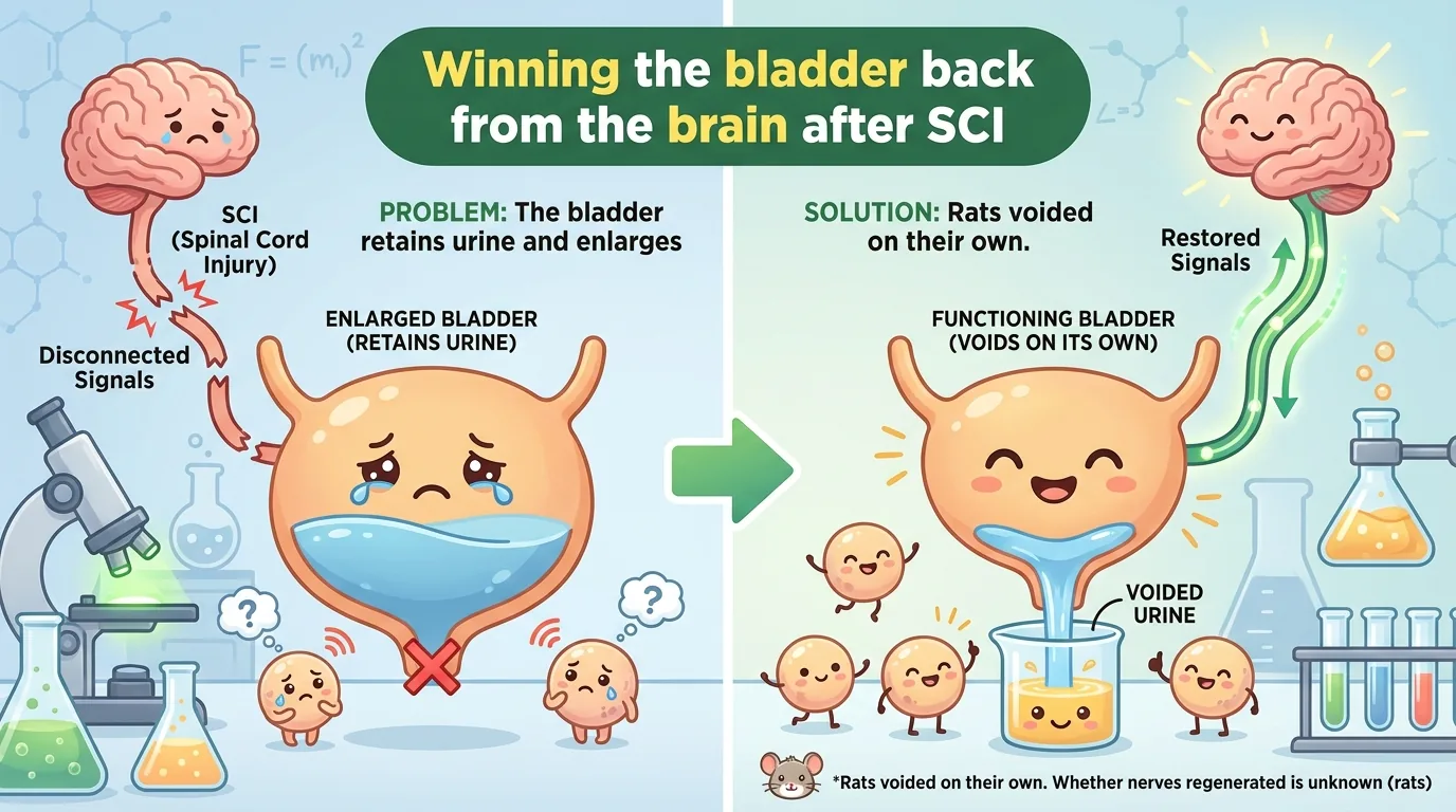
When people hear “spinal cord injury (SCI),” the first thing most of them picture is losing the ability to walk. Paralysis, a wheelchair, rehabilitation — that is usually as far as television and film go. But what actually troubles people with spinal cord injury most acutely, day after day, and what can even put their lives directly at risk, lies somewhere else entirely. It is that urine can no longer be passed at will.
More than 80% of people who sustain a spinal cord injury are said to live with disturbances of voiding and storage — what is known as neurogenic bladder. Even when urine collects in the bladder, no signal saying “this is the limit” reaches the brain, and conversely no command saying “you may go now” arrives from the brain. The accumulated urine refluxes toward the kidneys, infections recur, and if left alone it can lead to renal failure and death. Amid all the attention paid to recovery of walking, there is a reason patients themselves name the bladder as “the thing they most want fixed first.”
Ironically, though, this urgent problem has hardly been examined in preclinical spinal cord injury research (basic research using animals). There are countless assessments of motor function, yet experiments that measure the bladder carefully are astonishingly few. The paper introduced here cuts straight into precisely this “overlooked bladder.” It administers the clinical-stage anti-RGMa antibody elezanumab in a rat injury model that closely resembles human spinal trauma, and measures bladder function — and the neural plasticity behind it — with multiple techniques.
Let me say this clearly up front. This is a preclinical study conducted in female rats, not a story about bladders being cured in humans. Even so, a study that approaches the question “could a drug restore even a little of the brain–spinal cord circuitry that governs voiding?” from this many angles is valuable. Let us read through, in order, what was shown and what has not yet been shown.
Journal Information
- Paper title: Clinical stage anti-RGMa monoclonal antibody elezanumab promotes bladder recovery and neuroplasticity after traumatic spinal cord injury
- Authors: Andrea J. Mothe, Magdy Hassouna, Christopher Ahuja, Peer B. Jacobson, Lili Huang, Michael G. Fehlings, Philippe P. Monnier, Charles H. Tator
- Affiliations: Centered on the Krembil Brain Institute (Division of Experimental and Translational Neuroscience) at the University Health Network (UHN), University of Toronto, together with the University of Toronto Department of Surgery (Urology and Neurosurgery / Spine Program), the same university’s Department of Physiology, and the developer AbbVie (research sites in North Chicago, Illinois and Worcester, Massachusetts, USA)
- Journal: Biomedicine & Pharmacotherapy (published by Elsevier Masson SAS) / Volume 202 / article number 119769 / 2026
- DOI / link: 10.1016/j.biopha.2026.119769
- Impact Factor: roughly around 7.5 (the exact current value should be checked in Journal Citation Reports). Within its field it sits in Q1 (the top quartile) as a general pharmacology and medical journal.
- Open access: An open access paper. The license is CC BY-NC-ND 4.0 (Attribution-NonCommercial-NoDerivatives). © 2026 The Author(s), Published by Elsevier Masson SAS. Citation and sharing are permitted provided the source is stated, but there are restrictions on modification and commercial use (http://creativecommons.org/licenses/by-nc-nd/4.0/ ).
- Review timeline: Received March 15, 2026 / revised version received July 10, 2026 / accepted July 14, 2026 / published online July 21, 2026.
- Ethics approval: All procedures were reviewed and approved by the Animal Care Committee of the University Health Network research institute (AUP 2976) and complied with the policies of the Canadian Council on Animal Care. The animal experiments followed the ARRIVE guidelines. No human subjects were involved.
- Funding: Grants from Paralyzed Veterans of America, the Morton Cure Paralysis Fund, the Wings for Life Spinal Cord Research Foundation, the Krembil Foundation, and the University Health Network Foundation, plus in-kind support from AbbVie in the form of the antibody supply. The authors state that “the funders had no role in study design, data collection/analysis, the decision to publish, or manuscript preparation.”
- Conflicts of interest: The antibody elezanumab was developed by AbbVie, and it is disclosed that the inventor, Bernhard K. Mueller, currently holds all rights and commercial interests relating to the use of this antibody. Among the authors, L.H. and P.J. (the latter formerly) are/were AbbVie employees and may hold stock or similar interests. AbbVie provided no operating funds for this study, had no access to the data, and took no part in analysis or interpretation, but it is explicitly stated that AbbVie did participate in the review and approval of the manuscript.
- Data availability: “Data will be made available on request.” Supplementary data are posted with the online version.
Note that no explicit disclosure statement regarding the use of generative AI or large language models appears in the text (not applicable).
About the corresponding authors: Two people are designated as responsible (corresponding) authors in this paper — Dr. Andrea J. Mothe and Dr. Charles H. Tator (both are marked with an asterisk in the author list, and the UHN email addresses of both are given as contacts) — and both are based at the Krembil Brain Institute of UHN, University of Toronto. Dr. Charles H. Tator is a neurosurgeon who has been active in spinal cord injury research for a very long time. In 1974 he established Canada’s first acute spinal cord injury unit, and since then he has consistently studied the pathophysiology, prevention, and treatment of acute spinal cord injury and concussion. He is currently professor emeritus in the university’s Department of Surgery and emeritus senior scientist at the Krembil Brain Institute. He has received publicly verifiable honors such as Officer of the Order of Canada and induction into the Canadian Medical Hall of Fame. Dr. Andrea J. Mothe is a spinal cord injury researcher likewise based at the Krembil Brain Institute, who has spent years studying spinal cord regeneration through neural stem cell transplantation and the antibody therapy targeting RGMa that is central to this paper. Together with Dr. Tator, Dr. Philippe Monnier, and others, she has led a body of work on regeneration, plasticity, and repair after spinal cord injury using anti-RGMa human monoclonal antibodies. The co-author list also includes Dr. Michael G. Fehlings, well known in spinal surgery and regenerative medicine, as well as researchers from the developer AbbVie. Elezanumab is an antibody created by AbbVie (development code ABT-555).
What Was Not Known Until Now?
First, it is worth appreciating just how finely tuned an “orchestra of nerves” the act of voiding really is. When we casually relieve ourselves in the bathroom, a remarkably complex coordination is taking place among the brain, spinal cord, peripheral nerves, and bladder.
When urine collects in the bladder, that information travels through sensory nerves to the spinal cord and on to the brain. During storage, the sympathetic and somatic nerves act to “relax the bladder muscle (detrusor) and tighten the outlet of the urethra (external urethral sphincter, EUS).” Then, once the bladder is full, a command center centered on the pontine micturition centre (PMC) in the brainstem flips the switch, the parasympathetic nerves of the lumbosacral cord (L6-S1) contract the detrusor, and the sphincter relaxes at the same time — this exquisite timing is what makes smooth voiding possible.
This circuit involves not only the PMC but several brain regions, including the raphe nuclei and reticular nuclei of the brainstem, the locus coeruleus (LC), and the periaqueductal gray (PAG). It is an ensemble performed by “the conductor (the brain)” and “the players (the spinal cord and bladder)” as one.
👦 Student: So does that mean that when the spinal cord is damaged, the “communication cable connecting the conductor and the players” in that ensemble is severed?
🧬 Dr. Exotaro: Exactly so. In spinal cord injury, both the descending pathways from the brain and the local circuits of the spinal cord are cut off. Immediately after injury the bladder goes limp with “spinal shock,” and after a while reflexive voiding driven by the spinal cord alone begins. But this is a performance with no conductor, and the rhythm is all over the place. The bladder overfills, fails to empty, or contracts on its own. Overactivity, incontinence, residual urine — and the bladder itself enlarges.
The problem is that this state is not merely inconvenient. If urine accumulates while the pressure inside the bladder stays high, it refluxes to the kidneys, inviting renal damage and renal failure. Current management uses catheters, anticholinergic drugs, and botulinum toxin injections into the bladder, but all of these are symptomatic, many patients stop responding, and complication risks follow them around.
👦 Student: So there’s still no treatment that fundamentally “reconnects the severed cable.”
🧬 Dr. Exotaro: That is the biggest gap. And when you try to reconnect the cable, it turns out there is a “brake” inside the body that works to keep it from reconnecting. The leading example is today’s protagonist, RGMa.
RGMa (Repulsive Guidance Molecule a) is a protein that powerfully suppresses the extension of nerve processes (axons), and it is also involved in neuronal survival and differentiation. Originally it is a “repulsive signal” that guides axons in the right direction during development, but, awkwardly, when the central nervous system is injured by trauma or stroke, it increases excessively at the injury site. A variety of cells — from neurons to microglia to macrophages — release RGMa, and through the receptor neogenin they keep the brake pressed with the message “grow no further.” Axons that are trying hard to regenerate are blocked by this brake.
Put the other way around, if you release this brake called RGMa, might axonal regeneration and sprouting, neural sparing, and functional recovery be promoted? In rodent and monkey models of acute spinal cord injury, such effects have in fact been reported. And in the authors’ own earlier work, administering an antibody that neutralizes RGMa was shown to accelerate the recovery of spontaneous voiding after spinal cord injury. But exactly which urodynamic measures underpin that recovery, and what the neuroanatomical basis of the plasticity is — that remained unresolved. This paper sets out to fill the gap.
What Did This Paper Find?
The research team used adult female Wistar rats (270–300 g body weight, 66 in total). For the injury model, they adopted an “impact-compression” approach in which the spinal cord is clamped bilaterally with a modified aneurysm clip. A clip with a closing force of 21 g is applied at the T8-9 level of the thoracic cord for one minute, producing a moderate injury. Because an instantaneous impact is combined with sustained compression, it closely reproduces the mechanical pathophysiology of human spinal trauma, and the authors emphasize that one element of novelty is that this model has hardly ever been used in bladder research. Note that three animals were excluded after injury based on prespecified exclusion criteria.
The dosing protocol was as follows. Three hours after injury, a loading dose of 65 mg/kg of elezanumab (anti-RGMa antibody, RGMa Ab) was given intravenously (IV) via the tail vein, followed by 25 mg/kg weekly for six weeks (six doses in total). The control group received isotype control human IgG1 (hIgG) on the same schedule. Allocation was randomized using a computer-generated random sequence, and assessors measured under blinded conditions. Prespecifying the primary endpoints (spontaneous voiding score, urine retention volume, metabolic cage, cystometry, bladder and lesion histology, TH/5-HT quantification, BBB score, and so on) and then positioning retrograde/transsynaptic tracing and ELISA as secondary and exploratory items is methodologically honest. The behavioral assessments covered control n=16 and treated n=21.
Now let’s look at the results. First, motor function. The antibody-treated group showed significantly better recovery on the BBB locomotor score (0–21, higher is better) (at 6 weeks, control 9.5 vs treated 11.9, p=0.0257). The proportion reaching “BBB ≥10,” meaning weight-supported stepping, was 56% in controls versus 95% in the treated group (p=0.0118). On gait analysis (CatWalk), the regularity index was 85% in the treated group versus 54% in controls — here the effect size was large and the difference was clear-cut (p<0.0001).
And now to the main subject, the bladder. The proportion of rats that regained spontaneous voiding after injury was 95% in the treated group versus 37% in controls at six weeks. The urine retention volume, measured by daily manual bladder expression, was also significantly lower in the treated group. Looking at overnight (17-hour) voiding patterns in the metabolic cage, control rats showed an abnormal pattern of “fewer voids, with a larger volume per void.” In the treated group this voiding frequency was significantly higher than in controls (p=0.0264), the volume per void was smaller, and the pattern had returned closer to that of healthy rats. There was no between-group difference in water intake, so this cannot be explained by differences in how much they drank.
👦 Student: Why is “more voids with a smaller volume each time” the good direction? Frequent urination sounds like a bad thing to me.
🧬 Dr. Exotaro: Good question. The healthy state here is “little and often, and emptying properly.” The bladder after spinal cord injury is the opposite: “store and store, occasionally release a flood, and still not empty.” That very inability to empty becomes residual urine, raises the pressure, and is the root cause of kidney damage. So moving back toward voiding frequently and thoroughly makes sense as a way of protecting the kidneys.
The team went further and performed cystometry (bladder pressure measurement) in awake rats. Because anesthesia alters bladder behavior, they deliberately measured without it. Whereas healthy rats show regular contractions, the recordings from injured rats were disordered, with reflexive bladder activity and elevated internal pressure. Here the treated group had less reflexive activity and lower pressure than controls. At week 7 the mean intravesical pressure was 20.01 mmHg in the treated group versus 24.72 mmHg in controls (p=0.0475), and there was also a significant difference in the area under the pressure curve (AUC) (p=0.0274).
👦 Student: 20.01 versus 24.72 mmHg — that’s a difference of just under 5 mmHg, right? Is that a clinically meaningful difference?
🧬 Dr. Exotaro: You’ve put your finger on a sharp point. Looking at the numbers alone it seems “slight,” and p=0.0475 is right on the edge of 0.05. But in the bladder, sustained high internal pressure is itself what destroys the kidneys. So if the mean pressure falls and, at the same time, the disordered reflexive contractions settle down — a change in quality — it can mean more than the absolute difference suggests. That said, the authors themselves caution that in this experimental setup voiding contractions and non-voiding contractions cannot be reliably distinguished. So while you can say “the pressure went down,” there is not yet enough material to declare “voiding efficiency was fully improved.”
The shape of the bladder itself had changed too. The bladders of control rats had enlarged to roughly four times the healthy size, whereas in the treated group the enlargement stopped at about two-fold, significantly smaller. Bladder wet weight correlated with urine retention volume (Spearman r=0.54, p=0.0499 in the treated group), confirming the relationship that “the more urine is retained, the more the bladder hypertrophies.” However, it should be noted honestly that while bladder wall thickness was significantly greater in controls than in healthy animals (p=0.0416), there was no significant difference between the treated group and controls.
From here comes the most interesting part of this paper. It explores why the bladder improved from the side of neural connectivity, using three techniques.
The first is retrograde tracing. The spinal cord is transected at the L4 level and the fluorescent dye Fluoro-Gold (FG) is placed on the stump. Because the dye is taken up by cut axons and transported backward to the cell bodies, it lets you count “neurons in the brain whose axons had descended past the injury site” (n=4/group). In the treated group, FG-labeled neurons were significantly increased in several brain regions involved in voiding — the reticular nuclei (treated mean 3402 vs control 1960, p=0.0476, Hedges’ g=1.53), the PMC (860 vs 343, p=0.0093, g=2.31), and the locus coeruleus LC (1109 vs 244, p=0.0272, g=1.78). The red nucleus showed only a trend toward increase (p=0.0733, g=1.33), and the raphe nuclei, PAG, and sensorimotor cortex showed no significant differences. The authors themselves state explicitly that “these should be interpreted cautiously because of the small samples, parametric tests, and lack of correction for multiple comparisons.”
The second is transsynaptic tracing. Pseudorabies virus (PRV) is injected into the detrusor muscle of the bladder. Because this virus travels from neuron to neuron across synapses, it can visualize “how much circuitry connects the bladder to the brain” (uninjured n=7, SCI control n=5, SCI+RGMa Ab n=7). The result spoke to the severity of spinal cord injury. Whereas healthy rats show solid labeling in the brainstem, in control rats after injury only 1 of 5 animals showed any labeling in the brain at all. In the treated group there was a consistent trend toward more labeling, but the between-group difference was not statistically significant (p>0.05). With so few labeled cells to begin with, interpretation is limited — something the authors again admit honestly.
👦 Student: Retrograde tracing (FG) showed a significant increase, but transsynaptic tracing (PRV) showed no significant difference. They’re both looking at “connections,” so why do the conclusions disagree?
🧬 Dr. Exotaro: What they look at is subtly different. FG counts “neurons in the brain whose axons had descended past the injury site.” PRV, on the other hand, starts from the bladder muscle and only labels cells if it can climb several synapses all the way to the brain. In other words, PRV asks the stricter question of whether the bladder and brain are “truly connected as a functioning circuit.” The fact that in control rats only 1 of 5 reached the brain tells you how deep the disconnection is. So the increase in FG is encouraging, but it is too early to declare from that alone that “the bladder and brain have been correctly reconnected.” The very gap between the two techniques is the point that deserves careful reading in this study.
The third — and the one with the largest effect size — is the plasticity of descending fibres in the lumbosacral cord. The team quantified fibres positive for tyrosine hydroxylase (TH), a marker of catecholaminergic signaling, and for 5-HT (5-hydroxytryptamine), a marker of serotonergic signaling, in the spinal cord below the injury (control n=7, treated n=8). In control rats these fibres were almost absent below the injury site, the descending pathways having been severed. In the treated group, by contrast, the density of both TH and 5-HT fibres was markedly increased. In numbers, TH was significantly increased in the T10 ventral horn and the L1/2 intermediate gray matter (both p=0.0003, effect size r=1.00), and 5-HT in the T10 ventral horn (p=0.0006, r=0.96) and at L1/2 (p=0.0022, r=0.89), all with large effect sizes (Mann-Whitney test). Moreover, triple staining under confocal microscopy captured these TH-positive fibres forming synapses (synaptophysin-positive) in the vicinity of Onuf’s nucleus, where the motor neurons innervating the external urethral sphincter are clustered. It should be noted that this is the first study to examine the effect of RGMa inhibition on TH axonal plasticity.
👦 Student: So both the “cable” to the brain and the “wiring” inside the spinal cord had increased?
🧬 Dr. Exotaro: That is a fair summary. But I want to choose my words carefully. Whether the increased fibres represent the “sparing” of axons that were about to be cut, newly extended “sprouting,” or genuine “regeneration” — the authors themselves state that this incomplete injury model cannot distinguish among them. You can say that the wiring between conductor and players appears to have “thickened.” But whether that wiring is really delivering the right notes cannot be proven from this study alone.
The team also examined systemic inflammation on an exploratory basis. Measuring 27 cytokines/chemokines in week-7 plasma, the anti-inflammatory IL-10 was higher in the treated group (752.1 pg/ml vs 356.7 in controls, uncorrected p=0.0410), but significance disappeared once multiple-testing correction (FDR) was applied (q=0.5071). Other markers showed trends but no significant differences, so this cannot be called “proof of an anti-inflammatory effect”; it is a suggestive finding and nothing more. On histology, the treated group had a significantly higher rate of gray matter sparing at the lesion epicenter (p=0.021), but the authors interpret this by noting that “there was no association between intravesical pressure and tissue sparing, so tissue sparing is not the main driver of bladder recovery.”
How Will the Future Change? (The Road to the Clinic)
So how might these preclinical results connect to patients’ futures? The greatest significance is that they show the possibility that a single intervention, RGMa inhibition, might reach not only motor function but another urgent problem directly tied to survival: the recovery of bladder function. In spinal cord injury research, which has tended to skew toward walking, this study tackled that “overlooked priority” with a neuroregenerative approach and sought to substantiate the effect with cystometry and tracing rather than impressions — and that stance in itself throws a stone into the pond of future study design.
It also matters that elezanumab is a “clinical stage” antibody. This antibody has completed evaluation in a randomized, double-blind, placebo-controlled phase II trial (NCT04295538, AbbVie) in acute traumatic cervical spinal cord injury (AIS grade A/B, C4-7). It is a 52-week study in which dosing begins within 24 hours of injury and continues every four weeks through week 48; the primary endpoint is the change in upper extremity motor score, and the secondary endpoints include the self-care subscore of the Spinal Cord Independence Measure. In addition, an independent clinical trial in acute traumatic spinal cord injury using a different anti-RGMa antibody (NCT04683848, Mitsubishi) is also under way. This is a drug for which the “bridge” from bench to bedside is, at the level of clinical trials, already being built.
👦 Student: So can we hope that bladders will eventually be cured in humans too?
🧬 Dr. Exotaro: Here let me draw a firm line. What this paper showed is only “an effect on bladder function and neural plasticity in rats.” There is as yet no evidence anywhere that elezanumab restored bladder function in humans. In fact, to avoid confusion, there is something I should say honestly.
Elezanumab did not live up to expectations in human clinical trials in multiple sclerosis (MS). RADIUS-R, a phase II study in relapsing MS, and RADIUS-P in progressive MS had good safety and tolerability, but did not meet their primary efficacy endpoints — a fact this very paper states explicitly. The phase II study in acute spinal cord injury (NCT04295538) has completed, but the final efficacy results have not yet been published. A report that good safety was shown in a phase IIa study in acute ischemic stroke (NCT04309474) remains, at present, at the level of a conference abstract.
In short, elezanumab is “an investigational antibody that multiple human trials confirm can be given safely, but whose efficacy has not yet been proven.” That is exactly why the present bladder findings in rats are most accurately received not as proof that “it works in humans,” but as a proposal of a hypothesis and a direction — that “it is worth looking much more carefully at the bladder as an outcome in human trials.” If future clinical trials incorporate urodynamics and the quality of voiding into their secondary endpoints, and not just the upper extremity motor score, this preclinical study will serve as a signpost.
How to Read This Study Critically (Limitations, and the Road to Better Quality)
The better the study, the more honestly it discloses its limitations. This paper lists its own weaknesses almost as a model of how it should be done. First, the commendable points are plain: randomized allocation, blinded assessors, prespecified exclusion criteria and primary endpoints, ARRIVE compliance, and a priori power calculations with G*Power (histology n=8/group, functional assessment n=12/group for 80% power, α=0.05) — the methodological skeleton is solid. The fact that several independent techniques produced results pointing in the same direction is also more persuasive than a single-measure study. The novelty of assessing the bladder in a compression model that has rarely been used before stands out as well. On that basis, let’s look at the limitations.
First, only female rats were used. Taking into account sex differences in lower urinary tract physiology, in the anatomy of the external urethral sphincter and Onuf’s nucleus, and in hormonal regulation of voiding, the authors themselves state explicitly that “the findings may be sex-specific and require verification in males” (though they add that no sex differences were found in earlier work, and that efficacy has also been shown in male non-human primates). These results cannot be generalized as they stand to “humans” or to “men and women.”
Second, many of the p values sit just below 0.05. Intravesical pressure p=0.0475, BBB p=0.0257, the correlation between bladder weight and urine retention p=0.0499 — all significant, but none of them differences you could call “dramatic.” On the other hand, the gait regularity index and the TH/5-HT plasticity show large effect sizes. The fact that the strength of the effect varies from measure to measure should be accepted frankly.
Third, the limits of interpreting the tracing. Because the retrograde tracer FG was placed at L4, it captures the sympathetic circuits of L1-L2 but does not include the L6-S1 parasympathetic/external-urethral-sphincter circuits that are central to voiding. The regional comparisons of FG used small samples with no correction for multiplicity, and the authors themselves caution readers to “interpret carefully.” The transsynaptic PRV showed no significant between-group difference, only a trend, so interpretation is limited. And most fundamentally, “more supraspinal connections” and “those connections having functionally reinnervated the correct targets” are two different things. The authors also note that directly demonstrating the latter would require anterograde tracing from the PMC.
Fourth, central versus peripheral contributions cannot be separated. This study suggests central plasticity, but because RGMa expression in the bladder wall and elezanumab binding there were not examined, a peripheral contribution cannot be excluded. The effect may be central in origin, peripheral, or a combination of both.
Fifth, the handling of exploratory findings. The systemic anti-inflammatory effect, IL-10 included, lost significance after FDR correction. Because of technical constraints, cystometry could not reliably distinguish voiding from non-voiding contractions, and this measure was not included in the analysis. The development of anti-drug antibodies (ADA) against human IgG with repeated dosing was also not examined in this study.
So, how could this have been an even higher-quality study? Verification in both sexes to assess sex differences; extending the FG tracer down to L6-S1 to confirm reinnervation of the parasympathetic/sphincter circuits; direct proof of “arrival at functioning targets” through anterograde and intersectional tracing from the PMC; separating central from peripheral contributions by assessing RGMa expression and antibody binding in the bladder wall; and improving the robustness of p values hovering near 0.05 by increasing the sample size — these are the constructive “next moves.”
Dr. Exotaro’s Perspective
Having finished this paper, what resonated most strongly with my own interests was, in fact, not the bladder itself but the problem consciousness behind it: shining the light of neuroscience on an overlooked complication.
I too have worked on spinal cord injury and stroke using mesenchymal stem cells (MSCs) and their EVs (extracellular vesicles / exosomes). When we say “functional recovery,” our eyes are inevitably drawn to “can they walk,” yet what determines patients’ quality of life (QOL) and survival prognosis are the domains of voiding, defecation, pain, and autonomic function — “unglamorous but lethal,” so to speak. I straightforwardly sympathize with the stance of this study in putting the bladder, which patients name as their top priority, in the leading role, and in trying to measure it fully with cystometry and tracing rather than impressionistic argument.
What I found particularly interesting from a neuroscience standpoint is that the plasticity of the descending monoamine systems — serotonergic (5-HT) and catecholaminergic (TH) fibres — may hold the key to bladder recovery. This finding presses on us the view that recovery after spinal cord injury should be understood not as “rebuilding a single motor pathway” but as “reorganizing a network of multiple modulatory systems.” Since the repair we aim at with MSCs and EVs is likewise not the replacement of a single cell type or pathway but a push toward reorganizing the whole microenvironment and the whole network, there is common ground here.
But let me draw a calm line here as well. This study has nothing to do with exosome or mesenchymal stem cell therapy. It is an effect shown by a specific drug, an anti-RGMa antibody, in a preclinical model in female rats, and it shows nothing whatsoever about the efficacy of EV therapy. Its point of action also differs from the secretome-based therapies we work with. So I want to receive it not as a tailwind for my own research, but strictly as a suggestion from a neighboring field: that “with the bladder as the axis, the plasticity of descending monoamine systems could be a driver of recovery.” Whether it is an antibody that releases the brake called RGMa or an EV that tunes the microenvironment, the destination — giving a severed network room to connect once more — certainly overlaps.
Finally, one thing I want to emphasize. What makes this study genuinely valuable is not the brilliance of its conclusions. The p values hovering near 0.05, the transsynaptic labeling that fell short of significance, the anti-inflammatory finding that vanished under correction — the authors disclosed all of them without concealment. Being preclinical, being female-specific, being unable to show functional reinnervation directly — they made all of this explicit, and still carefully advanced the question of whether “the bladder might be reclaimed from the brain.” Hope in medicine can only ever be built on an accumulation of honest steps like these.