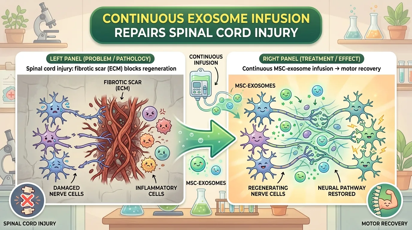
Some scientific questions need a device before they can be asked. Without a thermometer, “hot” stays a matter of opinion; without a clock, “fast” cannot be compared. Today’s question is of that type — does the same quantity of a drug work differently when it goes in all at once and when it seeps in over several days? The trouble is that a syringe is an instrument that fixes quantity and time together, and you cannot inject 4.6 micrograms “slowly.” The first thing this study did was choose a device.
What was infused were the small extracellular vesicles (sEVs) released by human bone marrow–derived mesenchymal stem cells (MSCs). This is the fraction commonly called “exosomes,” but isolating pure exosomes alone is difficult, and this paper follows the international guidance known as MISEV2023 in calling them sEVs.
Publication Details
- Paper link: doi:10.1016/j.neurot.2026.e00892
- Journal: Neurotherapeutics 23 (2026) e00892. An Original Article, accepted March 13, 2026. It is the official journal of the American Society for Experimental NeuroTherapeutics (ASENT) and, from the January 2024 issue, a fully open-access title published by Elsevier
- Impact Factor: 7.4 (2025 citation data). Note: this is the value listed by the third-party aggregator bioxbio; the original Clarivate JCR entry has not been checked
- License: CC BY 4.0. The full text can be read at PMC13054005
- Authors: Masahito Nakazaki, Karen L. Lankford, Masayuki Toyoshima, Yoshiaki Tanaka, Tomokazu S. Sumida, Jeffery D. Kocsis
- Funding and ethics: The RR&D and BLR&D services of the U.S. Department of Veterans Affairs (B7335R, B9260L). The animal experiments were approved by the IACUC of the VA Connecticut Healthcare System
Corresponding Author: Jeffery D. Kocsis
The corresponding author is Jeffery D. Kocsis. He is a professor of neurology and neuroscience at Yale University School of Medicine, associate director of the university’s Center for Neuroscience and Regeneration Research (CNRR), and a Senior Research Career Scientist with the U.S. Department of Veterans Affairs (Yale faculty profile). He took his PhD at Wayne State University in 1976 and trained as a postdoctoral fellow at Harvard Medical School and MIT. The CNRR introduces him as an expert in axonal regeneration and remyelination and in cell-based approaches to repairing nerve injury, and puts his output at more than 200 papers (CNRR administration page).
A single thread runs through that record. From the 1996 work in which transplanting Schwann cells into demyelinated adult rat spinal cord restored conduction properties to normal (Honmou O, Felts PA, Waxman SG, Kocsis JD. J Neurosci 1996;16(10):3199-3208), to the report that vesicles do not work unless they are given divided across three days (Nakazaki M, et al. J Extracell Vesicles 2021;10(11):e12137) — the present paper sits on the extension of a line running from cells to vesicles. The CNRR is located on the grounds of the Veterans Affairs hospital.
One further note: the collaboration with Sapporo Medical University that Yale announced in 2021 (intravenous autologous bone marrow–derived stem cells) was described in the release itself as a study in which “each patient served as his or her own control,” in which “the results were not blinded and no placebo control was used,” and which was “a preliminary, unblinded trial, and further study is needed to confirm the results” (Yale News, February 22, 2021) — and what went in there were cells, not vesicles.
What Wasn’t Understood Before?
Spinal Cord Injury in Japan Does Not Look the Way You Imagine
The picture most people carry of spinal cord injury usually sits at an angle to the reality. In a 2018 nationwide survey, the annual incidence of traumatic spinal cord injury in Japan was 49 cases per million population, the median age at injury was 70.0 years, cervical cord injuries accounted for 88.1%, and the commonest cause was not road traffic accidents but falls on level ground (38.6%) (Miyakoshi N, et al. Spinal Cord 2021;59(6):626-634). That is an entirely different face from the United States, where the mean age at injury is 44 and road traffic accidents come first. Recovery is scant as well: in the 2025 statistics from the U.S. National Spinal Cord Injury Statistical Center, the proportion of people reaching complete neurological recovery by discharge is “less than 1%” (NSCISC 2025 Facts and Figures).
The Accident Is an Instant; the Injury Goes On for Weeks
The injury does not end at the moment of impact. After the primary injury the blood–spinal cord barrier (BSCB) breaks down, and the leak spreads above and below the lesion over the course of six weeks (Matsushita T, et al. Exp Neurol 2015;267:152-164). Among the macrophages that pour in, the inflammatory M1 type dominates at first, and over days to weeks the balance tips toward the reparative M2 type. M2 cells clear away the debris and secrete extracellular matrix (ECM), building the scaffold along which axons grow. The problem lies past that point. When fibronectin and collagen I become excessive, the fibrous scar turns back into a barrier to regeneration. What matters is not excessive accumulation but balanced remodelling, the paper writes. Build the scaffolding too far and it becomes a wall.
👦 Student: Scar tissue really is the villain here, isn’t it?
🧬 Dr. Exotaro: It is not that simple. A 2016 paper in Nature reported that in adult mice, neither preventing astrocyte scar formation nor removing a scar that had already formed led severed axons to grow back on their own (Anderson MA, et al. Nature 2016;532:195-200). If anything, the scar appears to be on the side that helps regeneration. It is like the hoarding around a building site: taking it down is not what makes the work proceed. But the metaphor breaks. In the spinal cord the same molecule serves as scaffold and as wall. The target is not the hoarding but the fibrous ECM inside it.
There Was a Reason to Suspect “Time” Rather Than “Amount”
MSCs given intravenously are not found in the injured spinal cord. In the Exp Neurol 2015 study cited above, the administered MSCs were not detected in the cord; they collected mainly in the lungs and stayed there for a few days. The present paper cites this too, writing that intravenously delivered MSCs do not significantly target the lesion. They cannot be detected, and yet function recovers. The hypothesis offered to explain this is that the cells lodged in the lungs release vesicles into the bloodstream over time, and those vesicles travel to the lesion. And indeed, intravenously injected MSC-derived sEVs have been reported to accumulate selectively at the lesion and to be taken up by CD206-positive M2 macrophages (Lankford KL, et al. PLoS One 2018;13(1):e0190358).
And there is a strange asymmetry. The parent MSCs improved motor function after a single injection, whereas the same total amount of vesicles did nothing when given all at once, and only worked once it was divided across three days (covered previously on this site). If the total amount is the same and the outcome differs, then what is working is something other than amount.
👦 Student: If the amount is the same, surely it makes no difference whether you give it all at once or split it up?
🧬 Dr. Exotaro: The closest analogy is probably vitamin C. Swallow a large dose at once and the surplus simply leaves in the urine. There is a comparable observation for vesicles: the excess is cleared by the kidneys within 24 hours (the figure appears in the abstract, but the substance of it comes from earlier work). A low concentration is enough to change a cell, but it has to be maintained for several days — that is how the authors see it. But the metaphor breaks, because vesicles rewrite the gene expression of the cells that take them up. And this paper never once measured the vesicle concentration in the blood. “The concentration was maintained” is an inference.
So what happens if you simply never let it break off? A syringe cannot test that. If you tried to keep it unbroken by injecting once an hour — that is, 24 times a day — the rat would be restrained 24 times a day, and that stress would contaminate the result. The first thing this paper solved was this design problem.
What Did This Paper Find?
A Pump the Size of a Soybean, Driven by Salt Alone
The device used was the Alzet osmotic pump 1003D (DURECT Corporation). It has no battery and no motor: water enters by osmosis through the semipermeable membrane of the outer shell, and the swelling salt layer squeezes the reservoir and forces the contents out — it runs on nothing but the force that makes a dried pickled plum swell when you soak it in water. Overall length 1.5 cm, 1.0 μL per hour for three days: those are the manufacturer’s nominal values, and the same specification sheet also says “not for human use” (ALZET 1003D specification sheet). The paper itself does not state a flow rate, so these are nominal figures. The catheter runs from the external jugular vein toward the heart, the pump is implanted subcutaneously between the shoulder blades, and for the six-day schedule it is replaced after three days.
The identity of the vesicles is examined as well. Electron microscopy showed numerous vesicles 70–150 nm in diameter, and the mean particle size by nanoparticle tracking analysis (NTA) was 130 nm (n = 3 preparations). CD63, CD9 and Alix were enriched relative to the parent cells, and the endoplasmic reticulum marker calnexin was barely present. Holding the vesicles at 37 °C for three days to mimic residence inside the pump produced decreases of 11.7 ± 1.4% in mean particle size, 19.1 ± 4.1% in particle concentration and 1.47 ± 0.88% in total protein (n = 3 in each case). RNA yield, however, had fallen by 29.3 ± 1.16% (n = 3). The authors’ conclusion is that structural and molecular integrity was largely maintained (Are the vesicles from day 40 of culture the same product?).
The animals were mature male Sprague–Dawley rats (185–215 g). A contusion injury was made at the ninth thoracic vertebra with an Infinite Horizon impactor (the impact is given in the paper in two forms side by side, “22.5 N” and “225 kdyn”). Only 101 of the 340 rats that underwent contusion met the strict inclusion criteria — a Basso–Beattie–Bresnahan (BBB) locomotor score of 0 at three days after injury and 0.5 at seven days. Nine animals were lost to complications the authors judged unrelated to treatment (hindlimb infections associated with immobility after spinal cord injury, among others), leaving 92 for the behavioural analysis. In the breakdown the paper provides, though, seven of those nine were in PBS groups, and not one came from a continuously infused sEV group. Treatment began on day 7 after injury, observation ran to day 70, and there were eight groups. The daily injection groups received 1.53 μg per dose in 100 μL of PBS; the pump groups had 9.45 μg loaded into 100 μL and delivered 4.6 μg over three days (the paper does not explain the reason for the difference, and it is not for a reader to fill in).
| Group (hMSC-sEV) | Delivery | Total dose | BBB at day 70 |
|---|---|---|---|
| 3 days, daily injection | 1.53 μg × 3 doses | 4.6 μg (about 2.5 × 10⁹ particles) | 8.2 |
| 3 days, continuous infusion | Pump for 3 days | 4.6 μg (about 2.5 × 10⁹ particles) | 9.7 ± 0.7 |
| 6 days, daily injection | 1.53 μg × 6 doses | 9.2 μg (about 5.0 × 10⁹ particles) | 8.6 ± 0.9 |
| 6 days, continuous infusion | Pump for 6 days with one replacement | 9.2 μg (about 5.0 × 10⁹ particles) | 12.9 ± 0.9 |
“The same total dose” holds only for comparisons between groups run over the same number of days. In extending from three days to six, the Results section states plainly that the total dose of hMSC-sEV was doubled in both the daily injection and the pump groups. Read the table vertically and time and amount move together; only when read horizontally does the total dose match (for 8.2 the paper gives two versions, 8.2 ± 0.8 and 8.2 ± 0.7, so only the mean is listed here).
Doubling the Amount Bought Nothing
The three-day protocol. Both the daily injection and the continuous infusion sEV groups recovered significantly more than the PBS groups (P < 0.001 at every time point from day 14 to day 70). At day 70 the figures were 8.2 for daily injection and 9.7 ± 0.7 for continuous infusion (n = 15 each), against 3.8 ± 0.9 (n = 8) and 3.3 ± 0.9 (n = 7) for PBS. The two sEV groups did not differ significantly at day 70, however; continuous infusion came out ahead from day 14 to day 35. The paper’s own phrasing is that the time course of recovery was accelerated, while at the final time point it amounted only to a trend toward improvement.
The six-day protocol. Here the order of the findings matters. The total dose has been doubled. On top of that, two results. First, doubling the total dose while staying with daily injections changed recovery at no time point at all. Second, only the group in which the same 9.2 μg was infused continuously over six days improved. The six-day continuous infusion group scored significantly higher than the six-day daily injection group at every time point from day 14 to day 70, reaching 12.9 ± 0.9 at day 70. The main text does not give the significance level at each time point (the legend to Fig. 2 defines #p < 0.05, ##p < 0.01, ###p < 0.001 for the Daily-versus-Pump comparison), and the comparison that carries P < 0.001 is the one showing that the final score at day 70 was higher than under every other condition.
👦 Student: So 12.9 against 8.6 means a difference of about fifty percent?
🧬 Dr. Exotaro: That is a calculation you must not do. The BBB is an ordinal scale from 0 to 21, and its intervals are not equal. Going from 0 to 1 (slight movement of a joint) and going from 8 to 9 (from placing the sole of the foot down, to bearing weight on it) are the same single point, but they carry completely different weight. Incidentally, this yardstick also scores whether the tail is raised; a rat that cannot support itself on its hindlimbs drags its tail. A healthy rat scores 21. Looking at the graduations themselves, “occasional” forelimb–hindlimb coordination scores 12 and “frequent” coordination scores 13, so 12.9 falls between the two. But this is a group mean. From a mean you cannot read off how far any individual rat came to be able to walk.
sEV treatment also promoted recovery of body weight and body length (at day 70 of the three-day protocol, body weights were 295.9 ± 7.2 g and 305.3 ± 10.0 g in the PBS groups against 356.2 ± 6.6 g and 344.4 ± 7.0 g in the sEV groups, P < 0.05). But the sEV groups did not differ from one another, and the same held for crown–rump length. Growth is probably governed by a separate pathway (the hepatic growth hormone receptor and IGF-1), the authors reason, as earlier work covered here previously has already reported. If the BBB scores had risen because general condition was better, body weight ought to have moved in the same way; this dissociation makes it considerably harder to read the difference between delivery modes as nothing more than a difference in general condition. The absence of a difference in weight and length does not in itself prove the absence of confounding, but the fact remains that only motor function depended on how the vesicles were delivered.
Predicted 914, Measured 1,445, Overlap 101
The stage on which the mechanism is pursued is the culture dish. Monocytes from rat bone marrow were cultured for 11 days and polarised to the M2 type with IL-4 and IL-13 (the medium was at pH 6). Two days later, vesicles labelled with the fluorescent dye DiR were added together with a fraction enriched in central myelin for 48 hours, and the cells containing DiR were sorted out. The control was not PBS but vesicles from human serum (hS-sEV), a design that separates out the question of whether anything at all that happens to be a vesicle will work. That said, the statement that giving serum-derived sEVs to animals had no effect on recovery appears only as a single line in the text describing a preliminary experiment; the result is shown neither in a figure nor as a number.
The proportion of macrophages that took up vesicles was 70.8 ± 2.0% for hMSC-sEV and 37.0 ± 1.0% for hS-sEV (n = 3 per group, P < 0.001). This is the proportion of positive cells, not the amount taken up per cell. Sorting the DiR-positive cells and comparing them, 1,564 genes were up and 1,445 genes were down on the hMSC-sEV side relative to the hS-sEV side. In the pathway analysis, fibrosis, wound healing and ECM organisation all swung to the inhibited side, and what stood on the activated side was the IL-4/IL-13 used to induce the M2 phenotype.
Inside the vesicles, hMSC-sEV carried 119 miRs at increased levels and 113 at decreased levels compared with hS-sEV (n = 3 preparations each). Asking IPA (Ingenuity Pathway Analysis) to predict the candidate targets of the top 30 increased miRs returned 914 genes. Overlaying these on the 1,445 genes actually found to be down in cultured macrophages, 101 genes fell into both sets, and among them stand the collagens (Col1a1, Col5a1-3) and fibronectin (Fn1). 914 is prediction, 1,445 is measurement, and 101 is where the two overlap — they must not be read as one thing. Put the other way round, the remainder of the 914 simply did not enter the set of “decreased genes” that satisfies the strict criteria this analysis adopted (adjusted p value < 0.05 and an expression ratio below 1/4), which is not the same as ruling out the possibility that they fell a little. And there is no test of whether an overlap of 101 exceeds what chance would predict. One further note: the pathway names “hepatic fibrosis” and “idiopathic pulmonary fibrosis” are labels the analysis software attaches to gene sets, and do not mean that cirrhosis is occurring in the spinal cord.
In the Matched-Total-Dose Comparison, Only COL1A1 Fell
On immunostaining at 14 days after injury, fibronectin (FN1) deposition around the lesion in the PBS groups co-localised mainly with the M2 marker CD206, and was reduced in the six-day continuous infusion sEV group (no quantitative values are given). On western blot at 70 days after injury (n = 5 per group, n = 3 for the non-SCI control), FN1, COL1A1 and COL5A2 were all significantly lower in the sEV-treated groups than in any of the PBS groups. Here is the core of the paper: six days of continuous infusion lowered COL1A1 expression significantly more than six days of daily injection delivering the same total dose. In the comparison that holds total dose constant, COL1A1 was the only molecule in which a difference between delivery modes showed up; for FN1 and COL5A2 no difference is demonstrated.
👦 Student: So the more you reduce collagen, the better?
🧬 Dr. Exotaro: That is the interesting part, because the authors themselves write that it is not. Fibronectin is known to promote neurite outgrowth in peripheral nerve through β1 integrin, and a scaffold of type I collagen has been shown to support axonal growth in spinal cord injury models as well. The same molecule serves as scaffold and as wall. It is like wet concrete: while it is still flowing you can pour it into a form, but once it sets your only option is to break it. Fibronectin is thought to assemble from the soluble form into matrix at around day 7 after injury, which coincides with the day dosing began. But the authors do not write that this was their reason for the choice, and “it coincides” is as far as one can go. Incidentally, this pairs neatly — contents against delivery — with the study that followed what gets loaded into the vesicles.
How Will the Future Change? (The Path to the Clinic)
What this study moved is not the content of the treatment but the way the blueprint is written. The paper’s conclusion states that the temporal distribution of administration may be an important factor determining the efficacy of hMSC-sEV treatment for spinal cord injury. A review covered here previously left the optimal dose, number of administrations and route undetermined (Polishing MSC-EVs into drug delivery vehicles); this amounts to one preclinical answer coming back to that question. Six days in a rat, converted straight into hours, is 144 hours. Transposed to a person that would mean close to a week of continuous central venous infusion — but there is no established method for interspecies conversion of duration, so this is not even a rule of thumb.
👦 Student: Couldn’t you just keep a person hooked up for six days as well?
🧬 Dr. Exotaro: Technically, yes. For inpatients in the subacute phase, central venous catheters are in routine use. But there is a price. Catheter-related bloodstream infection, thrombosis, length of stay and cost. Over long durations and large volumes, the fraction of vesicles lost by adsorbing to the inner wall of the infusion line is not negligible either. And what was shown goes no further than “six days was better than three” — there is no nine-day or twelve-day group. On the dose side, the per-injection amount in the daily groups stayed fixed at 1.53 μg, and there is no group that held duration constant and varied only the amount. Dose conversion, too, gives answers that differ several-fold depending on whether you scale by body weight or by body surface area.
The question of the time window also remains. Treatment began in the subacute phase on day 7 after injury, and there is not one group in which dosing started in the chronic phase. What is more, the more correct this mechanism is — suppressing ECM production by actively working macrophages — the worse the outlook for the chronic phase becomes, because once the matrix has fully set and macrophage activity has quietened down, the very production that needs suppressing should already have diminished. This, though, is a prediction from mechanism, not a measured fact. The paper itself sees deposits of FN1 and COL1A1, partly co-localised with CD206-positive macrophages, at the lesion in the PBS groups 70 days after injury. Neither when nor by how much the target diminishes, nor what would happen if dosing began in the chronic phase, has been established. The only thing that is certain is the shape of the argument: the more correct the mechanism, the harder application to the chronic phase becomes.
As of September 2026, a search of ClinicalTrials.gov turned up no registered interventional trial of MSC-EV or exosome therapy for traumatic spinal cord injury. The closest entry in that registry, a phase 1 trial in syringomyelia (NCT07295067), is for a different disease and uses intrathecal rather than intravenous administration.
On the cell side, things are further along. In Japan a regenerative medicine product consisting of intravenous autologous bone marrow–derived MSCs received conditional and time-limited approval on December 28, 2018, and full approval was reported to have been applied for on November 19, 2025 (the outcome has not been confirmed). The evidence base, however, was a domestic phase II trial of 13 cases with no control group, and the PMDA review report itself records that “no control group was established, and the possibility cannot be ruled out that factors other than this product (spontaneous recovery, the influence of rehabilitation, and so on) affected the results … we consider the data obtained to be limited.” In a news article in Nature (Cyranoski D. Nature 2019;565:544-545), ten experts who spoke to the reporter pointed to the insufficiency of the evidence, and in that same article the corresponding author of the present paper commented that continued research would be needed to establish efficacy adequately. Under the same framework, HeartSheet was reported to have been refused approval on July 19, 2024, and Collategene to have had its application withdrawn. This framework is not a free pass.
And there is a distinction that must always be kept. In both the Japanese regenerative medicine product just described and the Yale clinical study, what went into humans were cells, not vesicles. It is not that vesicles have no human experience whatsoever: a phase 1 trial has been reported in which human umbilical cord MSC–derived exosomes were given intrathecally to nine patients with complete spinal cord injury in the subacute phase (Akhlaghpasand M, et al. Stem Cell Res Ther 2024;15(1):264). It did not come up in the search above because that trial is registered in Iran’s national registry (IRCT20200502047277N1) and not on ClinicalTrials.gov. Both the cell source and the route of administration are different from the present paper, though, and there is no human track record for a protocol of continuously infusing bone marrow MSC–derived sEVs intravenously. What the present paper administers are sEVs derived from human bone marrow MSCs (Lonza PT-2501), and the subjects are rats. Neither “clinical” nor “patient” appears anywhere in its text. A study that makes no claim to human application should not be wired up to the clinic on someone else’s initiative. Rather than a failing, that should be read as lying outside its range.
How to Critically Read This Research (Limitations and Paths to Improve It)
Let us grant the strengths first, and fairly. The design philosophy — hold the total dose constant and vary only the mode of administration — is the right one. Most preclinical EV papers stop at “EV versus saline”; this study built that comparison at both three and six days, and placed PBS controls on both the injection and the pump sides. The inclusion criteria are strict, and the ordering, with allocation falling on the day treatment starts, is correct as well. The characterisation goes as far as showing negative markers, meeting the minimum requirements of MISEV2023, and on the question of whether the vesicles degrade inside the pump, the authors go and plug the first hole a critic would prod at, themselves. Nor do they hide the limitations. Biodistribution was not measured; establishing causality for the miRs would require gain- and loss-of-function experiments; reducing ECM is not simply a good thing. All three of these are the authors’ own statements, and it would be a mistake to read them as blind spots the paper failed to notice.
Here are the points where a line should be drawn.
1. The reporting of blinding does not cover the most important comparison. Blinding is mentioned in exactly one place: that BBB scoring was done by someone who had not been told what was administered.
👦 Student: But surely you can tell just by looking whether there’s a pump in the animal’s back?
🧬 Dr. Exotaro: That is precisely the weak point. A device 1.5 cm long (the manufacturer’s figure) is implanted between the shoulder blades, and there is a surgical wound on the neck. The six-day pump groups also undergo replacement surgery on day 3 — a procedure that falls equally on the sEV and the PBS groups, but one that looks different from the daily injection groups. And injection versus pump is the very comparison at the core of this paper’s novelty. Yet the paper only writes that the scorer was not told what was administered; it describes neither any measure taken to make the mode of administration unrecognisable from appearance, nor the implantation of a dummy pump in the daily injection groups. Concealing the contents (PBS or sEV) is easy enough, but the reader has no way of confirming whether the mode of delivery was concealed as well. The fix is to implant a dummy pump in the daily injection groups too, and to say so. It is not difficult.
2. In “six days is better than three,” time and amount move together. Extending from three days to six also doubles the total dose (4.6 → 9.2 μg), so the difference between 12.9 and 9.7 cannot separate duration from dose. What the matched-total comparison — six-day continuous infusion 12.9 ± 0.9 against six-day daily injection 8.6 ± 0.9 — demonstrates is not duration itself but the effect of temporal distribution: over the same number of days and at the same total dose, the difference between giving it in boluses and letting it run. The abstract’s phrase “despite the same total dose” looks at odds with the main text if it is read as a comparison of three days against six, but this is not an error in the paper so much as a sentence that is easy to misconstrue. Even so, the internal evidence supporting the temporal-distribution account is there in the same paper. Staying with daily injections while extending from three days to six and doubling the total dose left recovery unchanged from the three-day daily injection group at every time point — and an explanation that says total dose is what determines efficacy cannot account for that result. There is no group in which the per-dose amount was raised, however, so what has been refuted is the axis of total dose, not dose-response in general.
3. There is a distance between “it worked” and “why it worked.” The chain the paper draws has five links: (1) continuous infusion → (2) M2 macrophages at the lesion take the vesicles up → (3) miRs suppress fibrosis-related mRNAs → (4) ECM decreases → (5) motor function recovers. What the data show are (4) and (5). Link (2) rests on a citation of earlier work, and the authors themselves write that biodistribution under continuous infusion was not measured. Link (3) is a correlation — an intersection of prediction and measurement — not a test of causality. As for (4) leading to (5), all that is said is that the extent of the decrease in these proteins correlated roughly with the extent of recovery; there is no correlation coefficient and no scatter plot.
The way (4) is measured calls for care as well. The western blot homogenises an 8 mm length of spinal cord centred on the lesion in its entirety, runs equal amounts of protein as determined by the BCA method, and quantifies against GAPDH as the internal standard. If more neural tissue survives in the treated groups, GAPDH increases and the ratio to ECM falls. Whether “ECM decreased” or “neural tissue survived and diluted it” cannot be told apart. The conclusion that treatment is beneficial does not change, but the claim about mechanism does.
4. There are holes in the reporting conventions that make the work impossible to check. The BBB is an ordinal scale, but the analysis is a two-way repeated-measures ANOVA with Sidak’s post hoc test — it is handled as an equal-interval continuous variable, and there is no statement that normality or sphericity was confirmed. Everything is reported against thresholds; there are no exact p values, no effect sizes and no confidence intervals. Error is given as SEM, so the sEV groups at around n = 15 look more precise than the PBS groups at n = 7–8. Randomisation is stated explicitly, but the method and the concealment of allocation are not written down, and the ARRIVE guidelines are cited in their 2010 version. Neither band quantification nor immunostaining carries any statement of blinding. This is a point about reporting standards, not an indictment.
5. There is no systematic safety evaluation anywhere. Unlike the preceding three, this is a point the authors do not list among their limitations. What is administered are human-derived vesicles, and what receives them are normal Sprague–Dawley rats with no mention of immunosuppression. And these are run into a vein continuously for up to six days. The text touching on safety extends as far as the nine deaths and euthanasias judged unrelated to treatment and their breakdown by group, and no further. Anti-human antibody titres, organ histology, blood chemistry — even the renal function whose relevance the paper itself invokes when it writes that surplus vesicles are cleared by the kidneys — show no sign of having been measured. Words corresponding to “safety,” “adverse,” “toxic” and “immunogen” do not appear anywhere in the main text or the supplementary material either (this is a statement about the presence of words). And, as seen above, extending from three days to six doubles the total dose (4.6 → 9.2 μg), so the total protein mass of human-derived sEV that the six-day groups receive is twice that of the three-day groups. Immunogenicity was not evaluated. The best-performing group is the six-day one.
And then, the very premise of “the same total dose” has never been measured. The pump was loaded with 9.45 μg, and what is said to be delivered is 4.6 μg over three days. The paper does not explain the reason for the gap, nor does it measure how many μg actually entered the bloodstream. The premise of the central comparison is an assumption, not a verified fact.
The reach of the conclusions is narrow too. Only male rats were used, and the words “female” and “sex” appear neither in the main text nor in the supplementary material. The injury model is a T9 contusion and nothing else, which is a different setting from the cervical injuries that make up 88.1% of traumatic spinal cord injury in Japan. As seen above, of the 340 rats that underwent contusion only 101 met the inclusion criteria, so nearly 70% dropped out at the door (92 remained for the behavioural analysis after a further nine were removed). This is a good design for raising homogeneity, but to exactly that extent the conclusions become a story about a narrow subpopulation of uniform severity. What was measured was the locomotor score and body weight and length, and nothing else: bladder function, pain, spasticity, autonomic function and sensation were none of them assessed (in the text “bladder” refers to postoperative manual expression care, and “pain” only to the description of analgesics).
The culture experiments, too, run under artificial conditions — myelin fragments added to a medium adjusted to pH 6 in order to maximise uptake — and the gene counts of 1,564 and 1,445 sit on top of that. (How the replicates were counted is not clear either. The Methods state that bone marrow monocytes from six rats were divided across three conditions, which is two animals per condition, while the uptake proportions in the Results are given as n = 3. How the two correspond, and whether this n = 3 represents biological replicates at the level of animals, is not written.) No GEO or SRA accession number is given for the sequencing data; data availability extends only to a statement that it is available from the corresponding author on reasonable request. From the text one cannot reach where the public data sit. A third party cannot check the arithmetic of the mechanism section on the spot.
How could this have been settled? Four measures, cheapest first. (1) Implant a dummy pump in the daily injection groups too, and write that you did — the reader would then be able to confirm that blinding held for the mode of delivery as well. (2) Add a group that runs for six days but at the same total dose as three days, 4.6 μg — if the effect matched six-day continuous infusion, duration was doing the work; if it matched three-day continuous infusion, total dose was (although this group halves the flow rate, so duration and rate would still move together). (3) Plot band density against BBB score for each individual animal — this could be done right now with the data already in hand, and it would put a number on the arrow from (4) to (5). (4) Quantify the fluid left in the pumps, deposit the sequencing data in GEO or SRA and give the accession number — “the same total dose” would become a fact, and the mechanism section would become verifiable.
One last note: although RNA yield fell by 29.3 ± 1.16% over the three days that simulated conditions inside the pump, the authors conclude that structural and molecular integrity was largely maintained. In a paper that argues the miRs are the leading players, that reading deserved to be dug into a little further.
Dr. Exotaro’s Perspective
As a physician specialising in regenerative medicine, I have long taken an interest in research applying MSC-derived extracellular vesicles to neurological disease. Reading from that position, what stayed with me longest was not the number 12.9 but the fact the paper passes over in a single line: that doubling the amount did nothing at all.
If it does not work, give more. In clinical practice that is the most natural move there is. That it fails here suggests the point of action of this treatment lies not with a receptor that saturates with concentration but with a cell state that gets rewritten over time. If so, the protagonist of a treatment protocol is not the dose but the length of time over which delivery goes unbroken. What a hospital would have to provide changes too — infusion pumps, line management, stability of the preparation inside a reservoir, several days of admission. The medical resources required are not those of a drug you give once and are done with.
To talk about this in clinic, one has to keep three distances in mind.
The first is the distance of scale. There is no conversion table between the rat BBB score and the human ASIA Impairment Scale. Nobody can translate “the BBB rose by 4 points” into “the AIS improved by one grade.”
The second is the distance of time. The rats in this study could be started on treatment on day 7 after injury. That the same thing would happen in the spinal cord of someone two years out from injury is hard for me to picture, because the more correct this paper’s explanation is, the more the ECM production that needs suppressing should have quietened down by the chronic phase. But there is no experiment in this paper that checked that. There is no group started in the chronic phase. Without being able to say either that it works or that it does not, the mechanism alone tells us to be cautious. As a physician I cannot leave that suspension vague and hand over hope alone.
The third is the distance of the functions that were measured. A rat being able to walk is not the same thing as a person being able to get to the toilet unaided. This study measured neither bladder nor pain.
And this is a preclinical rat study. When the U.S. NINDS tried to replicate promising spinal cord injury treatments at independent laboratories, many of the experiments could not be reproduced (Steward O, et al. Exp Neurol 2012;233(2):597-605). This is not a criticism of the present paper; it is the rule of the field.
Even so, what I value in this paper is that it puts the result that did not work squarely on the page. That doubling the total dose produced no additional gain must have been an awkward result to write. Having left it in rather than erasing it, the paper carries through the argument that what worked was not increasing the amount but not letting it break off. Entering from the device makes the position of this study clear. It is not a study that found a new molecule; it is a study that changed one thing only — how the vesicles already in hand are handed over — and measured again. Whether a substance becomes a drug is, I think, usually decided by unglamorous work of exactly this kind.
This article was written with the assistance of AI.