
Extracellular vesicles (EVs) are tiny sac-like structures secreted by cells, and several subtypes exist. These subtypes are distinguished by size, biological properties, biogenesis mechanism, and function. Below, the major classes of extracellular vesicles are described.
Exosomes
Exosomes are extracellular vesicles with a diameter of about 30–150 nm, secreted from the multivesicular bodies (MVBs) within the cytoplasm. Exosomes contain proteins, lipids, and nucleic acids (mRNA, miRNA, lncRNA, etc.), and by transporting these biological molecules to target cells they promote intercellular communication. Exosomes are involved in many biological processes, including the immune response, neurotransmission, formation of the tumor microenvironment, and cellular senescence and apoptosis.
Ectosomes
Ectosomes are small membrane-coated particles that cells release into the environment. These particles are usually formed from the cell surface by “blebbing” or “pinching” (that is, a process in which a portion of the cell membrane separates to form a new small particle). Ectosomes can carry a variety of molecules from the original cell, and as a result can transmit information to other cells.
Ectosomes are widely recognized as being involved in intercellular communication. They contain biologically important substances such as proteins, lipids, RNA, and DNA. These components may differ depending on how the ectosomes are formed and from which cells they are released. This allows ectosomes to function as “packages” for conveying specific information to specific cells.
Ectosomes may also be useful for the diagnosis and treatment of disease. For example, certain cancer cells are known to release ectosomes containing specific proteins. By detecting these ectosomes, physicians may be able to identify the presence or progression of cancer. Furthermore, ectosomes may serve as “vectors” or “transport vehicles” carrying drugs. This could become a new way of delivering drugs to specific cells.
What is the difference from exosomes?
Both exosomes and ectosomes are small membrane-coated particles released by cells, but there are differences in their process of formation and their roles.
Exosomes are formed through a process in which an intracellular structure called the endosome matures and forms small vesicles (intraluminal vesicles) inside the cell. When these endosomes fuse with the cell membrane, these vesicles are released outside the cell, and these are exosomes. Exosomes often play the role of carrying various substances—proteins, lipids, RNA, and even DNA—for transmitting information between cells.
Ectosomes, on the other hand, are formed by separating directly from the cell surface. This process is called “blebbing” or “pinching” (that is, a process in which a portion of the cell membrane separates to form a new small particle). Ectosomes also play a role in information transmission, but their contents and the mechanism of their release from the cell differ from those of exosomes.
Meldolesi J. Exosomes and Ectosomes in Intercellular Communication. Curr Biol. 2018 Apr 23;28(8):R435-R444. doi: 10.1016/j.cub.2018.01.059. PMID: 29689228.
Exomere
Exomeres are a type of extracellular particle that are smaller in size compared with exosomes and microvesicles. This term arose from recent research seeking to understand the diversity and complexity of extracellular particles.
Exomeres are about 35 nanometers in diameter, considerably smaller than exosomes (usually 40–100 nanometers in diameter) and microvesicles (which have diameters of several hundred nanometers).
Research has shown that exomeres carry their own distinct set of biological molecules—namely, proteins, lipids, and nucleic acids. The unique composition of exomeres distinguishes them from other types of extracellular particles and suggests that they have their own biological functions.
Nation GK, Saffold CE, Pua HH. Secret messengers: Extracellular RNA communication in the immune system. Immunol Rev. 2021 Nov;304(1):62-76. doi: 10.1111/imr.13027. Epub 2021 Sep 20. PMID: 34542176; PMCID: PMC8756459.
Microvesicles
Microvesicles are extracellular vesicles with a diameter of about 100–1000 nm, generated by direct budding from the cell membrane. Microvesicles contain biological molecules such as lipids, proteins, and nucleic acids, and, like exosomes, are involved in intercellular communication. Microvesicles are involved in diverse biological processes, including the immune response, coagulation reactions, and the proliferation, invasion, and metastasis of tumor cells.
Apoptotic bodies
Apoptotic bodies are extracellular vesicles with a diameter of about 500–5000 nm, generated during the process of apoptosis (programmed cell death). Apoptotic bodies contain proteins and nucleic acids involved in apoptosis, and they promote the phagocytosis of the remnants of apoptotic cells by neighboring cells. This enables the efficient removal of the remnants of apoptotic cells and the suppression of inflammatory responses. Apoptotic bodies play an important role in maintaining tissue homeostasis and in regulating the immune system.
Oncosomes
Oncosomes are large extracellular vesicles derived from cancer cells, with a diameter of about 1000–10000 nm. Oncosomes contain proteins, nucleic acids, lipids, and other components involved in the proliferation, invasion, and metastasis of cancer cells. It has been suggested that these biological molecules affect surrounding normal cells and immune cells and promote the progression of cancer.
Exopher
Exophers are a type of membrane-bound extracellular vesicle (EV) released from cells into the external space. Exophers are notable for their size, being on average large—about 4 microns in diameter—and have the ability to expel intact organelles such as mitochondria and lysosomes as cargo. Exophers have been confirmed to be released from the neurons and muscles of nematodes, and even from the cardiomyocytes of mice. Exophers may remain connected to the cell that originally produced them by membranous filaments resembling tunneling nanotubes. Exophers share similarities with large oncosomes, but they differ in that they are produced from physiologically normal cells rather than from tumor-associated abnormal cells.
The production of exophers is thought to be a mechanism by which cells maintain homeostasis. Exophers are produced in response to intracellular protein aggregation, reactive oxygen species (ROS), heat, high-osmotic environments, starvation, and even spaceflight. The production of exophers has been found to depend on extracellular receptor signaling. Specifically, the production of exophers in nematodes involves two MAPK pathways, namely epidermal growth factor (EGF) and fibroblast growth factor (FGF) signaling. Furthermore, the extracellular signaling receptor MERTK expressed by cardiac-resident macrophages is required for the phagocytic clearance of exophers in mouse-derived cardiac tissue.
Exophers may be associated with disease. In the mouse heart, removing macrophages or blocking their ability to take up exophers causes inflammation and ventricular dysregulation. Furthermore, exophers may promote the spread of pathological proteins in neurodegenerative diseases through their ability to carry aggregated proteins, including the human huntingtin protein, to the exterior of neurons.
Turek M, Banasiak K, Piechota M, Shanmugam N, Macias M, Śliwińska MA, Niklewicz M, Kowalski K, Nowak N, Chacinska A, Pokrzywa W. Muscle-derived exophers promote reproductive fitness. EMBO Rep. 2021 Aug 4;22(8):e52071. doi: 10.15252/embr.202052071. Epub 2021 Jul 20. PMID: 34288362; PMCID: PMC8339713.
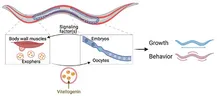
Muscle-derived exophers promote reproductive fitness - EMBO Reports
Other Extracellular Vesicles
Extracellular vesicles are classified into various subtypes based on size and biological properties, but as research advances, new subtypes may be discovered. In addition, extracellular vesicles can display different characteristics depending on cell type and state, and it has been reported that tissue-specific extracellular vesicles and disease-specific extracellular vesicles exist.
Future Directions
In the latest version, MISEV2018, it is recommended that, rather than the names “exosome” and “microvesicle,” extracellular vesicles be classified by size, with those of 200 nm or less called “small extracellular vesicles” (sEVs) and those of 200 nm or more called “medium/large EVs” (m/l EVs).
In addition, for the tetraspanins (CD9, CD63, CD81)—a transmembrane protein family that is highly expressed on extracellular vesicles and known as exosome markers—because expression levels vary depending on cell type, recovery method, and size, it is also recommended that notations such as (CD63+/CD81+-EVs) be added.
The classification of extracellular vesicles is expected to become even more detailed as research advances. By establishing standard classifications and isolation/purification methods for extracellular vesicles, the reproducibility and comparison of research results will become easier, and a deeper understanding of the biological properties and functions of extracellular vesicles is anticipated. Moreover, it is expected to contribute to the development of diagnostic and therapeutic methods using extracellular vesicles and to the elucidation of the mechanisms of disease onset and progression.
References
- Raposo, G., & Stoorvogel, W. (2013). Extracellular vesicles: Exosomes, microvesicles, and friends. The Journal of Cell Biology, 200(4), 373-383. This paper reviews the classification, biological functions, and in vivo roles of extracellular vesicles.
- Tkach, M., & Théry, C. (2016). Communication by extracellular vesicles: Where we are and where we need to go. Cell, 164(6), 1226-1232. A review focusing on intercellular communication by extracellular vesicles, explaining the current state of extracellular vesicle research and future challenges.
- Colombo, M., Raposo, G., & Théry, C. (2014). Biogenesis, secretion, and intercellular interactions of exosomes and other extracellular vesicles. Annual review of cell and developmental biology, 30, 255-289. A comprehensive review of the biogenesis, secretion, and intercellular interactions of extracellular vesicles, explaining in detail the biological processes of extracellular vesicles, including exosomes.
- Maas, S. L., Breakefield, X. O., & Weaver, A. M. (2017). Extracellular vesicles: Unique intercellular delivery vehicles. Trends in cell biology, 27(3), 172-188. A review focusing on the fact that extracellular vesicles provide a unique means of transport in intercellular communication, explaining the mechanisms of the generation, transport, and uptake of extracellular vesicles.