Journal Information
- Article link: 10.1002/jev2.702
- Journal: Journal of Extracellular Vesicles
- Impact Factor: approximately 25 (estimated value for 2023)
- About the journal: The Journal of Extracellular Vesicles (JEV) is one of the most authoritative academic journals in this field, specializing in research on extracellular vesicles (EVs). It publishes a wide range of cutting-edge research findings on the biology, function, and clinical applications of EVs.
Summary
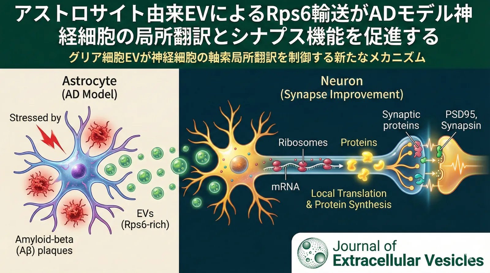
This study elucidates a mechanism by which astrocyte-secreted exosomes (EVs) regulate local translation in neuronal axons and improve synaptic function in an experimental model of Alzheimer’s disease (AD). Astrocytes exposed to amyloid β (Aβ) secrete EVs rich in a ribosomal protein called Rps6, and as these EVs are transported to neuronal axons, local protein synthesis is promoted. This finding reveals a previously unknown communication mechanism in which glial cells regulate local translation in neurons via EVs, bringing a new perspective to our understanding of AD pathology.
Background of the Study
Alzheimer’s disease (AD) is a neurodegenerative disorder characterized by cognitive decline, and its pathology is deeply involved in neuronal dysfunction and synaptic loss. In recent years, the role of not only neurons but also glial cells, particularly astrocytes, has attracted attention in AD pathology. Astrocytes are the most abundant glial cells in the brain and have diverse functions, including supporting neurons, regulating ions and neurotransmitters, and controlling synapse formation. Astrocytes are also known to secrete extracellular vesicles called exosomes (EVs). EVs contain various molecules such as proteins, nucleic acids, and lipids, and are thought to mediate intercellular communication.
Owing to their morphological complexity and high degree of compartmentalization, neurons are highly dependent on the local synthesis and transport of proteins. Traditionally, neuronal proteins were thought to be synthesized in the cell body and transported to distal sites such as axons and dendrites. However, in recent years, a mechanism in which mRNA is transported to distal sites and translated locally has attracted attention. This local translation is thought to play an important role in synaptic plasticity and neural circuit formation, but many aspects of its regulatory mechanisms remain unclear. In particular, whether glial cells regulate local translation in neurons via EVs had hardly been studied.
In this study, focusing on the possibility that astrocyte-secreted EVs regulate local translation in neuronal axons and improve synaptic function in an AD model, the researchers analyzed this mechanism in detail.
Introduction to the Authors and Laboratory
The corresponding author of this paper is Dr. Eva Maria Valente, and the final author is Dr. Stefania Gribaudo.
Dr. Eva Maria Valente is a researcher affiliated with the Fondazione IRCCS Istituto Neurologico Carlo Besta (Carlo Besta Neurological Institute) in Italy. Her laboratory focuses on elucidating the molecular mechanisms of hereditary neurological diseases, particularly movement disorders such as Parkinson’s disease. Through research using genetic screening, cell biology, and animal models, her research group investigates the identification of gene mutations that cause these diseases and the effects they have on neuronal function. Specifically, they analyze the role of genes in intracellular processes such as autophagy, mitochondrial dysfunction, and protein aggregation, aiming to elucidate disease pathology. She is particularly known for her research on LRRK2, a gene responsible for familial Parkinson’s disease, and has published many papers on the regulation of LRRK2 kinase activity and the effects of its mutations on neuronal survival and function.
Dr. Stefania Gribaudo’s laboratory is also affiliated with the Fondazione IRCCS Istituto Neurologico Carlo Besta and conducts research focusing on the role of glial cells in neurodegenerative diseases, particularly Alzheimer’s disease. Detailed information about the laboratory’s website and specific research content is currently not easily available online. However, from their published papers, it is evident that they are investigating how glial cells such as astrocytes and microglia are involved in pathological processes such as inflammation, oxidative stress, and protein aggregation. It is also thought that they are pursuing research with an interest in how communication between glial cells and neurons via extracellular vesicles (EVs) affects the progression of neurodegenerative diseases.
As background leading up to this study, Dr. Valente’s laboratory has long been engaged in elucidating the molecular mechanisms of neurodegenerative diseases, focusing in particular on the functional analysis of genes responsible for hereditary Parkinson’s disease. Meanwhile, Dr. Gribaudo’s laboratory has focused on the role of glial cells in Alzheimer’s disease and has advanced research on intercellular communication via EVs. It is thought that this time, the two laboratories jointly analyzed the effects of astrocyte-derived EVs on local translation in neurons, thereby revealing a new pathological mechanism of AD. This research suggests that crosstalk between glial cells and neurons may play an important role in the progression of neurodegenerative diseases, and it may open new avenues for the development of future AD therapies.
Main Findings
Overview of the Experimental System and Animal Model
In this study, an experimental system was constructed using primary cultured neurons and astrocytes to investigate the effects of astrocyte-derived exosomes (EVs) exposed to amyloid β (Aβ) on local translation in neuronal axons.
- Cell culture: Primary cultured neurons were prepared from mouse cortex and cultured under conditions that promote neuronal survival and differentiation. Astrocytes were similarly primary-cultured from mouse cortex and, by exposure to Aβ oligomers (Aβ42), a state mimicking AD pathology was created.
- Exosome isolation: EVs were isolated from the culture medium of Aβ-exposed or non-exposed astrocytes using ultracentrifugation. The isolated EVs were confirmed to be exosomes by verifying their size, shape, and expression of protein markers.
- Co-culture experiments: The isolated astrocyte-derived EVs were added to the neuronal culture medium and incubated for a certain period. Subsequently, changes in protein synthesis in neuronal axons, the amount of synapse-related proteins, and structural changes in synapses were evaluated.
Elucidation of the Molecular Mechanism
- Identification of Rps6: Proteomic analysis revealed that astrocyte-derived EVs exposed to Aβ were rich in the ribosomal protein Rps6. Rps6 is a constituent protein of the 40S subunit of the ribosome and plays an important role in the initiation and regulation of protein translation. Western blotting confirmed the increase of Rps6 in Aβ-exposed astrocyte-derived EVs.
- Experimental method details: Proteins were extracted from EVs isolated from astrocytes, and proteomic analysis was performed by mass spectrometry. The proteins contained in the EVs were identified by matching the obtained peptide sequences against a database. The presence of Rps6 was confirmed by Western blotting using a specific antibody. EVs and cell extracts were separated by SDS-PAGE, transferred to a PVDF membrane, and blotted with an anti-Rps6 antibody. Proteins were detected using an HRP-labeled secondary antibody, and the bands were visualized by chemiluminescence.
- Transport of Rps6 to axons: Fluorescently labeled Rps6 protein or Rps6 mRNA was encapsulated in EVs, and co-culture experiments with neurons were performed. Confocal microscopy confirmed that EV-derived Rps6 was transported to neuronal axons.
- Experimental method details: Rps6 protein was labeled with Alexa Fluor dye and introduced into EVs. Alternatively, Rps6 mRNA was introduced into EVs using a lipofection reagent. These EVs were co-cultured with neurons and observed by confocal microscopy after a certain period. Immunostaining of β-tubulin, an axonal marker, was performed to identify the localization of Rps6.
- Promotion of local translation: When Aβ-exposed astrocyte-derived EVs were added to neurons, protein synthesis in the axons increased significantly. This increase in protein synthesis was suppressed by knockdown of Rps6. A puromycin incorporation assay was used to quantitatively evaluate the synthesis of nascent proteins.
- Experimental method details: Neurons were co-cultured with Aβ-exposed astrocyte-derived EVs, and puromycin was added in the presence or absence of cycloheximide, a protein synthesis inhibitor. Puromycin binds to tRNA, is incorporated into the ribosome, and is incorporated into the polypeptide chain, thereby halting protein synthesis. Nascent proteins incorporating puromycin were detected by immunostaining using an anti-puromycin antibody. The amount of protein synthesis was quantitatively evaluated by measuring fluorescence intensity.
- Improvement of synaptic function: When Aβ-exposed astrocyte-derived EVs were added to neurons, the expression of synapse-related proteins (synapsin, PSD95) increased, and the structural integrity of synapses improved. Electrophysiological evaluation confirmed an improvement in synaptic transmission efficiency.
- Experimental method details: Neurons were co-cultured with Aβ-exposed astrocyte-derived EVs, and the amount of synapse-related proteins was measured by Western blotting. In addition, morphological changes in synapses were observed by electron microscopy. The electrophysiological function of synapses was evaluated using the patch-clamp method. Neurons were configured in the whole-cell configuration, the membrane potential was clamped, and postsynaptic currents were recorded. Synaptic transmission efficiency was evaluated by measuring the frequency and amplitude of excitatory postsynaptic currents (EPSCs).
- Phosphorylation of Rps6: Rps6 is known to be phosphorylated by S6 kinase (S6K). When Aβ-exposed astrocyte-derived EVs were added to neurons, the phosphorylation level of Rps6 increased. Administration of an S6K inhibitor suppressed the phosphorylation of Rps6 and the increase in protein synthesis in the axons.
- Experimental method details: Neurons were co-cultured with Aβ-exposed astrocyte-derived EVs, and the phosphorylation level of Rps6 was measured by Western blotting using a phosphorylation-specific antibody. PF-4708671, an S6K inhibitor, was administered to neurons, and its effect on Rps6 phosphorylation and protein synthesis was evaluated. The effect of the inhibitor was judged by whether the level of phosphorylated Rps6 decreased compared to the absence of the inhibitor.
Details of Cellular Responses
- Cell-type-specific changes: This study uses two types of cells: neurons and astrocytes. The changes due to Aβ exposure were mainly observed in astrocytes, and a mechanism is suggested in which these changes are transmitted to neurons via EVs. Using immunostaining and flow cytometry, changes in the expression of specific markers in each cell type were analyzed.
- Experimental method details: After fixing the cells, they were reacted with a primary antibody against a neuronal marker (NeuN) or an astrocyte marker (GFAP). Each cell type was visualized using a fluorescently labeled secondary antibody. Images were captured by confocal microscopy, and the fluorescence intensity of each marker was quantitatively measured. In flow cytometry, cells were made into a single-cell suspension by trypsinization and reacted with fluorescently labeled antibodies. The cells were analyzed with a flow cytometer, and the expression level of each marker was quantitatively evaluated.
- Dynamics of intracellular organelles: With the uptake of EVs, changes were observed in the dynamics of intracellular organelles such as endosomes and lysosomes in neuronal axons. Live-cell imaging revealed that EVs were taken up by neurons via endocytosis and transported through the endosomal pathway.
- Experimental method details: Fluorescent dyes (e.g., DiO, DiI) were inserted into the EV membrane to prepare fluorescently labeled EVs. Neurons and fluorescently labeled EVs were co-cultured and observed by time-lapse confocal microscopy. Using neurons expressing fluorescent proteins bound to marker proteins of endosomes or lysosomes, the uptake and intracellular transport of EVs were observed simultaneously. Analysis of time-lapse images traced the process by which EVs were taken up into endosomes and transported to lysosomes.
- Mechanism of cell fate determination: Activation of Rps6 may affect cell fates such as neuronal survival, axonal growth, and synapse formation. In this study, how the activation of Rps6 affects these cell fates was analyzed in detail. The role of Rps6 was evaluated using cell viability assays, axon elongation assays, synapse formation assays, and others.
- Experimental method details: In the cell viability assay, neurons were co-cultured with Aβ-exposed astrocyte-derived EVs, and cell viability was measured after a certain period. Cell viability was evaluated using the MTT assay or the LDH assay. In the axon elongation assay, neurons were seeded on Matrigel-coated culture dishes, and Aβ-exposed astrocyte-derived EVs were added. After a certain period, the length of the axons was measured, and axon elongation was quantitatively evaluated. In the synapse formation assay, neurons were cultured at high density to promote synapse formation. Aβ-exposed astrocyte-derived EVs were added, and the number of synapses was evaluated by immunostaining for the co-localization of synapsin and PSD95. Images were captured by confocal microscopy, and the number of synapses was quantitatively measured.
Integrative Understanding at the Tissue Level
This study is based on cell culture experiments, but its findings have important implications for understanding the interaction between neurons and glial cells at the tissue level, particularly in brain tissue. In the future, it is expected that experiments using AD model animals will verify the findings of this study at the tissue level.
- Changes in tissue structure: This study suggests that the structural integrity of synapses is improved. Since synaptic loss is observed in the brain tissue of AD patients, the findings of this study may lead to the development of new therapeutic strategies that suppress the progression of AD pathology. Changes in tissue structure were analyzed in detail through observation of the fine structure of synapses by electron microscopy and quantitative evaluation of synaptic marker proteins by immunohistochemistry.
- Experimental method details: Brain tissue of AD model mice was collected, fixed, and then paraffin-embedded or resin-embedded. Paraffin-embedded tissue was sectioned and then subjected to HE staining or immunohistochemical staining. Resin-embedded tissue was ultra-thin sectioned and then observed by electron microscopy. In immunohistochemical staining, antibodies against synaptic marker proteins (synapsin, PSD95) were used to quantitatively evaluate the number of synapses. In electron microscopy observation, the fine structure of synapses (such as the width of the synaptic cleft and the number of synaptic vesicles) was analyzed in detail.
- Effects on tissue function: Improvement of synaptic function leads to improved neurotransmission efficiency and may contribute to the improvement of cognitive function. Electrophysiological evaluation has confirmed an improvement in synaptic transmission efficiency. It is important to evaluate the effects on cognitive function through behavioral experiments using AD model animals (e.g., the Morris water maze test, the Y-maze test).
- Experimental method details: Using AD model mice, the Morris water maze test or the Y-maze test was performed. In the Morris water maze test, mice were guided to a platform in a water tank and made to learn the position of the platform. After learning, the platform was removed, and how accurately the mice remembered the position of the platform was evaluated. In the Y-maze test, mice were placed in a Y-shaped maze, and the number of entries into a novel arm was measured. The greater the number of entries into the novel arm, the higher the spatial cognitive ability was judged to be.
Validation Results in Animal Models
In this study, experiments using AD model mice were conducted to verify the results of the cell culture experiments. When Aβ-exposed astrocyte-derived EVs were administered intracerebroventricularly to AD model mice, the expression of synapse-related proteins increased, and the structural integrity of synapses improved. In addition, improvements in learning and memory abilities were observed in a cognitive function test (the Morris water maze test).
- Experimental method details: AD model mice (e.g., APP/PS1 mice) were administered Aβ-exposed astrocyte-derived EVs or PBS (control group) intracerebroventricularly. After a certain period, brain tissue was collected, and the amount of synapse-related proteins was measured by Western blotting. In addition, morphological changes in synapses were observed by electron microscopy. Cognitive function was evaluated using the Morris water maze test or the Y-maze test.
Discussion from a Specialized Perspective
Anti-aging
This study suggests that astrocyte-derived EVs may improve neuronal synaptic function, which is interesting also from an anti-aging perspective. It is known that with aging, the function of glial cells in the brain changes, and the support of neurons declines. Treatment using astrocyte-derived EVs may become a new strategy to prevent or improve the decline in cognitive function associated with aging. In particular, administration of EVs rich in Rps6 is expected to promote protein synthesis in neurons and support the maintenance and repair of synapses.
Regenerative Medicine (MSC / EV)
EVs derived from mesenchymal stem cells (MSCs) have been reported to have therapeutic effects against various diseases and are attracting attention in the field of regenerative medicine. This study shows that astrocyte-derived EVs bring beneficial effects to neurons, suggesting their potential as a new treatment for neurodegenerative diseases, similar to MSC-derived EVs. In particular, astrocyte-derived EVs are adapted to the microenvironment in the brain and may have higher targeting specificity than MSC-derived EVs. In the future, it is expected that the characteristics of astrocyte-derived EVs will be analyzed in more detail to develop optimal EV therapies for neurodegenerative diseases.
Nerve–Organ Interaction
This study focuses on the interaction between glial cells and neurons in the brain, but the nervous system is also closely linked with other organs, and the concept of nerve–organ interaction is important. For example, the gut microbiota is known to affect brain function, which is called the gut–brain axis. Astrocyte-derived EVs may be affected by changes in the gut microbiota, and these effects may be transmitted to neurons. In the future, research that clarifies how astrocyte-derived EVs affect the health of the whole body via nerve–organ interaction is important.
Future Perspectives
This study has revealed a new mechanism in which astrocyte-derived EVs regulate local translation in neurons and improve synaptic function, and it is expected to have a major impact on future AD research. Going forward, it is important to further advance research on the following points.
- Identification of EV cargo: It is necessary to analyze in detail how EV cargo other than Rps6 affects local translation in neurons. Using proteomic analysis, RNA sequencing, and so on, it is important to comprehensively identify the types of proteins and nucleic acids contained in EVs and to perform a functional analysis of each.
- Identification of target cells: It is necessary to clarify whether astrocyte-derived EVs act selectively on specific neuronal subtypes. Using single-cell RNA sequencing and so on, it is important to identify the target cells of EVs and to analyze the mechanism of action of EVs in those cells.
- Therapeutic application: Further research is needed toward the development of AD therapies using astrocyte-derived EVs. It is important to optimize the administration method, dosage, and timing of EV administration, and to verify the therapeutic effect in AD model animals. In addition, the safety and long-term effects of EVs must be carefully evaluated.
Conclusion
This study revealed that astrocyte-derived EVs promote local translation in neuronal axons via Rps6 and improve synaptic function. This finding suggests a new mechanism in which glial cells regulate neuronal function via EVs, and it may open new avenues for understanding AD pathology and developing therapies. In the future, it is expected that, by addressing challenges such as the identification of EV cargo, the identification of target cells, and therapeutic application, the development of AD therapies using astrocyte-derived EVs will be pursued.