Journal Information
- Article link: 10.1002/jev2.70109
- Journal: Journal of Extracellular Vesicles
- Impact Factor: approximately 25 (estimated value)
- About the journal: The Journal of Extracellular Vesicles (JEV) is the official journal of the International Society for Extracellular Vesicles (ISEV) and specializes in research on extracellular vesicles (EVs). It publishes high-quality papers covering every aspect of EV research, including EV biology, EV engineering, and EV-based diagnostics and therapeutics, and is recognized as one of the top journals in this field.
Summary
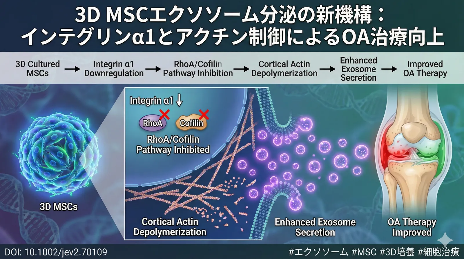
This study aimed to elucidate, at the molecular level, the mechanism by which three-dimensional (3D) culture of mesenchymal stem cells (MSCs) promotes secretion of extracellular vesicles (sEVs) compared with two-dimensional (2D) culture. The results revealed that in 3D-cultured MSCs, downregulation of integrin α1 (ITGA1) suppresses the RhoA/cofilin signaling pathway, thereby inducing depolymerization of cortical actin and promoting the release of sEVs. Furthermore, sEVs derived from 3D-cultured MSCs were shown to enhance therapeutic efficacy both in vitro and in vivo in rat models of osteoarthritis (OA) and wound healing. This study identifies RhoA/cofilin pathway-dependent cortical actin depolymerization as a novel mechanism that promotes sEV secretion, and provides new insights toward optimizing the yield and therapeutic efficacy of stem cell-derived sEVs.
1. What exactly is “cortical actin”?
Just inside the cell membrane, protein fibers called “actin filaments” are arranged in a mesh-like network. This is referred to as **cortical actin**.
- Role: It serves as the “framework” that maintains the cell’s shape, and also as a “physical barrier” that prevents substances inside the cell from leaking out at will.
2. What is “depolymerization”?
The breaking apart of fibers that have been polymerized (turned into a polymer) is called depolymerization. In other words, it is a state in which the tightly assembled mesh of actin fibers is broken down and the mesh loosens into a sparse arrangement.
3. Why is this important for “exosome secretion”?
Normally, exosomes (and the vesicles that contain them) inside the cell attempt to exit the cell membrane, but the mesh of cortical actin gets in the way and prevents them from easily getting out.
However, when **“cortical actin depolymerization”** occurs:
- Loss of the barrier: The obstructing mesh is broken down and disappears.
- Promotion of fusion: Vesicles containing exosomes can now reach the cell membrane smoothly and fuse with it.
- Increased secretion: As a result, large amounts of exosomes are released to the outside of the cell.
Background
Extracellular vesicles (EVs) are important mediators of intercellular communication, and they influence physiological and pathological processes by transporting bioactive substances such as proteins, nucleic acids, and lipids to target cells. In particular, extracellular vesicles (sEVs) derived from mesenchymal stem cells (MSCs) have been reported to exhibit a variety of therapeutic effects, including tissue repair, immunomodulation, and anti-inflammatory action, and have attracted considerable attention in the field of regenerative medicine. However, the amount of sEV secretion from MSCs is limited, and the development of effective methods to increase their yield is needed.
Compared with conventional two-dimensional (2D) culture, three-dimensional (3D) culture systems can bring cell morphology, gene expression, and cell-cell interactions closer to a more physiological state, which has been suggested to lead to improved cell function and enhanced sEV secretion. However, the molecular mechanism by which 3D culture promotes sEV secretion from MSCs has not been fully elucidated. This study aims to reveal a novel mechanism in which cortical actin depolymerization promotes sEV secretion in 3D-cultured MSCs, and—by analyzing this molecular mechanism in detail—to establish a foundation for optimizing the yield and therapeutic efficacy of sEVs.
Lab & Authors
This paper was published by the laboratory of Professor Lim Chwee Teck at the National University of Singapore (NUS).
About the Laboratory
Professor Lim Chwee Teck is affiliated with the Department of Biomedical Engineering and the Department of Mechanical Engineering at the National University of Singapore, and is also a Principal Investigator at the Mechanobiology Institute (MBI). The Lim laboratory focuses on the role of mechanical signaling at the cellular and tissue levels, and in particular studies the importance of mechanical forces in cancer, vascular disease, and stem cell biology. The laboratory employs a diverse array of technologies—including microfluidic devices, biomaterials, and imaging techniques—to investigate how cells sense and respond to mechanical stimuli. Its research findings to date have been published in leading journals such as Nature Materials, Nature Cell Biology, and PNAS. The present paper can be said to be research in line with one of the laboratory’s longstanding themes: the effect of cytoskeletal changes in 3D cell culture on exosome secretion.
The Lim laboratory was established in 1999 and has since produced numerous researchers. According to the laboratory’s website (https://me.nus.edu.sg/bme/people/academic-staff/lim-chwee-teck/), the laboratory’s main research themes are as follows.
- Mechanical properties of cells and the cytoskeleton: The laboratory studies how mechanical properties such as cell stiffness, viscoelasticity, and deformability affect cell function. In particular, it focuses on the mechanisms by which cytoskeletal components such as actin, myosin, and intermediate filaments control the mechanical properties of cells.
- Cell–extracellular matrix interactions: The extracellular matrix (ECM) functions not only as a scaffold for cells but also as an important signaling molecule that controls cell survival, proliferation, and differentiation. The laboratory investigates how cells sense and respond to the mechanical properties and composition of the ECM. In particular, it focuses on cell–ECM interactions mediated by cell adhesion molecules such as integrins.
- Cell manipulation using microfluidic devices: Because microfluidic devices can precisely control minute volumes of liquid, they are a powerful tool in cell biology research. The laboratory develops technologies that use microfluidic devices to measure the mechanical properties of cells, to apply mechanical stimuli to cells, and to separate and concentrate cells.
- Cancer mechanobiology: Cancer cells are known to exhibit mechanical properties that differ from those of normal cells. The laboratory studies how the mechanical properties of cancer cells—such as stiffness, deformability, and invasiveness—are involved in cancer metastasis and drug resistance. It also aims to develop new therapies that target the mechanical properties of cancer cells using microfluidic devices.
- Stem cell mechanobiology: Stem cells are special cells with the capacity for self-renewal and multipotency, and are expected to find applications in regenerative medicine. The laboratory studies the mechanical properties of stem cells and the mechanisms by which stem cells differentiate in response to mechanical stimuli. It also aims to develop technologies that use microfluidic devices to control stem cell differentiation.
The present paper can be said to be research in line with one of the laboratory’s longstanding themes: the effect of cytoskeletal changes in 3D cell culture on exosome secretion. In particular, it is considered to focus on how interactions with the extracellular matrix and the mechanical properties of cells affect exosome secretion.
About the Authors
The corresponding author of this paper is Professor Lim Chwee Teck. As noted above, Professor Lim is a renowned researcher at the National University of Singapore and one of the leaders in the field of bioengineering. His research combines cell mechanics, microfluidics, and biomedical applications, and focuses in particular on the study of cancer, stem cells, and extracellular vesicles. Professor Lim has received numerous awards for his notable contributions to the field. For example, he received the President’s Science and Technology Award of Singapore in 2016.
Professor Lim has published more than 400 papers to date, and his achievements are highly regarded internationally. His research has had a major impact on the fields of cell biology, biomaterials, and regenerative medicine, and his continued contributions are anticipated.
Background Leading to This Study
The Lim laboratory has, for many years, studied the relationship between the mechanical properties of cells and cell function. In particular, it has focused on how interactions with the extracellular matrix (ECM) and the mechanical stress placed on cells affect cell survival, proliferation, differentiation, and the secretion of extracellular vesicles. As part of this work, the present study focuses on the effect of 3D cell culture on the mechanical properties of cells and analyzes in detail how this affects exosome secretion. Considering that 3D cell culture is closer to a physiological state than 2D cell culture, the laboratory has long emphasized research on cell function using 3D cell culture. The present study is one of the results of that effort, and it is considered to have very important significance in that it elucidated, at the molecular level, the mechanism by which 3D cell culture promotes exosome secretion.
Key Findings – Molecular, Cellular, and Tissue Levels
Overview of the Experimental Systems and Animal Models
- Cells used: Human bone marrow-derived mesenchymal stem cells (hMSCs)
- Culture conditions: 2D culture (standard tissue culture flask) and 3D culture (Alvetex® scaffold). Both cultures used DMEM medium supplemented with 10% FBS and 1% penicillin/streptomycin. For 3D culture, cells were seeded onto an Alvetex® scaffold and cultured in the same medium as the 2D culture.
- Osteoarthritis (OA) model: OA was induced in 6-week-old male Sprague-Dawley rats by injecting monoiodoacetate (MIA) into the right knee joint. The MIA dose was 2 mg/50 μL.
- Wound healing model: A full-thickness skin defect 8 mm in diameter was created on the back of 8-week-old male Sprague-Dawley rats.
- sEV administration: In the OA model and the wound healing model, sEVs derived from 3D-cultured hMSCs (100 μg/50 μL PBS) were administered locally to the lesion site.
- Sample size: n = 5–8 per experimental group.
1. Detailed Explanation at the Molecular Level
1.1 Downregulation of Integrin α1 (ITGA1)
In 3D-cultured hMSCs, expression of integrin α1 (ITGA1) was significantly reduced compared with 2D culture, as confirmed by Western blotting (Figure 1A). ITGA1 is a cell surface receptor that binds to components of the extracellular matrix (ECM) such as collagen and laminin, and plays an important role in cell functions such as cell adhesion, cell migration, and cell differentiation. In the 3D culture environment, cells are thought to alter their interactions with the ECM and suppress ITGA1 expression, thereby inducing reorganization of the cytoskeleton. Specifically, cells were lysed, proteins were extracted, separated by SDS-PAGE electrophoresis, and transferred to a PVDF membrane. Detection was performed using a primary antibody against ITGA1 (e.g., Abcam ab30393) and an HRP-labeled secondary antibody. Bands were visualized by chemiluminescence and quantitatively analyzed with ImageJ software. ITGA1 expression levels were normalized to a housekeeping gene (e.g., GAPDH).
1.2 Suppression of the RhoA/Cofilin Signaling Pathway
Downregulation of ITGA1 causes suppression of the RhoA/cofilin signaling pathway. RhoA is a type of small GTPase involved in the contraction of the actin cytoskeleton and the regulation of cell adhesion. Cofilin is a protein that depolymerizes actin filaments; it is phosphorylated by ROCK (Rho-associated kinase), which is activated by RhoA, and its activity is thereby regulated. In 3D-cultured hMSCs, the phosphorylation levels of RhoA and cofilin were significantly reduced, as confirmed by Western blotting (Figure 1B, C). This suggests that downregulation of ITGA1 suppresses the activation of RhoA, which in turn reduces the phosphorylation of cofilin and promotes the depolymerization of actin filaments. Activation of RhoA can be assessed by measuring the amount of GTP-bound RhoA. GTP-bound RhoA is separated from the cell lysate by a pull-down assay and detected by Western blotting. Phosphorylation of cofilin is detected using a phosphorylated-cofilin-specific antibody.
1.3 Promotion of Actin Depolymerization
Suppression of the RhoA/cofilin signaling pathway promotes actin depolymerization. Actin is a major component of the cytoskeleton and is involved in various cell functions such as maintenance of cell morphology, cell motility, and intracellular transport. Actin exists in two forms—globular G-actin and filamentous F-actin—and enables dynamic reorganization of the cytoskeleton by repeatedly undergoing polymerization and depolymerization. In 3D-cultured hMSCs, the amount of F-actin decreased and the amount of G-actin increased, as confirmed by fluorescence microscopy and Western blotting (Figure 2A, B). This suggests that suppression of the RhoA/cofilin signaling pathway promotes the depolymerization of actin filaments, making the cytoskeleton more flexible. Actin polymerization is evaluated using fluorescently labeled phalloidin. Because phalloidin binds specifically to F-actin, observation by fluorescence microscopy makes it possible to evaluate the distribution and amount of F-actin. It is also possible to lyse the cells, separate G-actin and F-actin, and quantify the amount of each by Western blotting.
1.4 Suppression of sEV Secretion by Rab27A/B Knockdown
Rab27A and Rab27B are types of small GTPase involved in intracellular vesicle transport, particularly the secretion of extracellular vesicles (EVs). These proteins are known to regulate the process by which EVs fuse with the cell membrane and are released to the outside of the cell. In this study, hMSCs with Rab27A and Rab27B knocked down were created, and their effect on sEV secretion was examined. The results revealed that knockdown of Rab27A and Rab27B significantly suppressed sEV secretion (Figure 3A). Specifically, knockdown of Rab27A reduced sEV secretion to about 0.5-fold, and knockdown of Rab27B reduced sEV secretion to about 0.1-fold. This result suggests that Rab27A and Rab27B play essential roles in sEV secretion from hMSCs. Knockdown of Rab27A/B is performed using siRNA or shRNA. siRNA or shRNA is introduced into hMSCs to suppress the expression of Rab27A/B. The efficiency of knockdown is confirmed by qRT-PCR or Western blotting.
1.5 Changes in Rab27A/B Expression Levels in 2D/3D Culture
Interestingly, no significant difference was observed in the expression levels of Rab27A and Rab27B between 2D culture and 3D culture (Figure 3B). This suggests that the promotion of sEV secretion by 3D culture is not due to changes in the expression levels of Rab27A and Rab27B. Rather, the cortical actin depolymerization induced by 3D culture is thought to promote sEV secretion through a pathway independent of Rab27A/B. In qRT-PCR, the amount of Rab27A/B mRNA is measured. Total RNA is extracted, reverse-transcribed into cDNA, and PCR is performed using Rab27A/B-specific primers. Expression levels are normalized to a housekeeping gene (e.g., GAPDH). In Western blotting, the amount of protein expression is measured using antibodies against Rab27A/B.
2. Detailed Explanation at the Cellular Level
2.1 Morphological Changes in hMSCs
3D-cultured hMSCs were confirmed by phase-contrast microscopy to exhibit a more nearly spherical morphology compared with 2D culture (Figure 4A). This suggests that in the 3D culture environment, cells weaken their adhesion to the scaffold and reorganize their cytoskeleton, thereby adopting a freer morphology. Cell morphology is observed using phase-contrast microscopy or confocal laser microscopy. After fixing the cells, the cell membrane and cytoskeleton are stained and observed under a microscope. In 3D culture, it is also possible to observe cells infiltrating into the interior of the scaffold.
2.2 Reorganization of the Actin Cytoskeleton
In 3D-cultured hMSCs, the actin cytoskeleton was confirmed by fluorescence microscopy to be reorganized more flexibly (Figure 4B). Whereas in 2D culture the actin filaments are arranged in parallel along the cell membrane, in 3D culture the actin filaments were observed to be arranged more randomly and distributed uniformly throughout the cell. This suggests that the cortical actin depolymerization induced by 3D culture increases the flexibility of the cytoskeleton and promotes changes in cell morphology and cell motility. Visualization of the actin cytoskeleton is performed using fluorescently labeled phalloidin. Because phalloidin binds specifically to F-actin, observation by fluorescence microscopy allows the distribution and structure of actin filaments to be observed in detail.
2.3 Increase in the Amount of sEV Released
3D-cultured hMSCs were confirmed by nanoparticle tracking analysis (NTA) to release a significantly increased amount of sEVs compared with 2D culture (Figure 5A). NTA is a technique for measuring the size and concentration of microparticles (such as sEVs) in a liquid, and is widely used in sEV research. The fact that 3D culture increased the amount of sEVs released by approximately 2-fold suggests that 3D culture is an effective means of improving the production efficiency of sEVs. The amount of sEVs released is measured by collecting the culture supernatant, removing cells and debris by centrifugation, and then measuring it by NTA. In NTA, laser light is applied and the scattered light from the microparticles is analyzed to calculate the size and concentration of the particles.
2.4 Changes in the Uptake of sEVs
sEVs are taken up by target cells through mechanisms such as endocytosis, and they mediate intercellular communication by transporting bioactive substances—such as the proteins and nucleic acids contained within them—to the target cells. sEVs derived from 3D-cultured hMSCs were confirmed by fluorescence microscopy and flow cytometry to be taken up in increased amounts by target cells (e.g., chondrocytes and fibroblasts) compared with sEVs derived from 2D-cultured hMSCs (Figure 5B). This result suggests that 3D culture enhances the cellular uptake capacity of sEVs, which may contribute to improving the therapeutic efficacy of sEVs. The uptake capacity of sEVs is evaluated using fluorescently labeled sEVs. sEVs are labeled with a fluorescent dye (e.g., DiI or CFSE), added to target cells, and cultured for a certain period of time. The cells are then washed, and the amount of sEVs taken up into the cells is measured by fluorescence microscopy or flow cytometry.
3. Detailed Explanation at the Tissue Level
3.1 Cartilage-Protective Effect in the Osteoarthritis (OA) Model
Osteoarthritis (OA) is a chronic joint disease characterized by degeneration and destruction of the articular cartilage, and is one of the factors that markedly reduce the quality of life (QOL) of elderly people. In this study, when sEVs derived from 3D-cultured hMSCs were administered to an OA rat model, cartilage destruction was suppressed and the survival rate of chondrocytes improved, as confirmed by histological analysis (hematoxylin and eosin staining, safranin O staining, and toluidine blue staining) (Figure 6A, B). These results suggest that sEVs derived from 3D-cultured hMSCs have an effect of suppressing the progression of OA and protecting cartilage. In histological analysis, the joint tissue is excised, embedded in paraffin, sectioned into thin slices, and subjected to various stains. Hematoxylin and eosin staining allows observation of cell morphology and tissue structure. Safranin O staining and toluidine blue staining allow evaluation of the amount of cartilage proteoglycan. The degree of cartilage destruction is quantified using evaluation criteria such as the OARSI score.
3.2 Wound-Closure-Promoting Effect in the Wound Healing Model
Wound healing is a complex process that repairs skin damage, progressing through stages such as inflammation, cell proliferation, and tissue remodeling. In this study, when sEVs derived from 3D-cultured hMSCs were administered to a wound healing rat model, wound closure was promoted and the formation of granulation tissue was promoted, as confirmed by macroscopic observation and histological analysis (hematoxylin and eosin staining and Masson’s trichrome staining) (Figure 7A, B). These results suggest that sEVs derived from 3D-cultured hMSCs have an effect of promoting wound healing. The degree of wound closure is evaluated by measuring the wound area. The formation of granulation tissue is observed by hematoxylin and eosin staining, and the degree of angiogenesis and the amount of collagen deposition are evaluated. Masson’s trichrome staining allows detailed observation of the distribution of collagen.
4. Detailed Explanation of the Verification Results in Animal Models
4.1 Osteoarthritis (OA) Model
- Animal model: 6-week-old male Sprague-Dawley rats
- OA induction: Injection of monoiodoacetate (MIA) into the right knee joint (2 mg/50 μL)
- sEV administration: sEVs derived from 3D-cultured hMSCs (100 μg/50 μL PBS) administered into the knee joint cavity twice a week, four times in total
- Evaluation: Histological analysis (hematoxylin and eosin staining, safranin O staining, toluidine blue staining) performed at 4 weeks after administration
- Results: In the sEV-administered group, cartilage destruction due to MIA administration was significantly suppressed, and the survival rate of chondrocytes improved
- Statistical analysis: ANOVA followed by Tukey’s post-hoc test
4.2 Wound Healing Model
- Animal model: 8-week-old male Sprague-Dawley rats
- Wound creation: A full-thickness skin defect 8 mm in diameter created on the back
- sEV administration: sEVs derived from 3D-cultured hMSCs (100 μg/50 μL PBS) administered locally to the wound site
- Evaluation: The wound closure rate was measured at 14 days after administration, and histological analysis (hematoxylin and eosin staining, Masson’s trichrome staining) was performed
- Results: In the sEV-administered group, wound closure was significantly promoted, and the formation of granulation tissue was promoted
- Statistical analysis: ANOVA followed by Tukey’s post-hoc test
5. Specific Interpretation of the Experimental Data
5.1 Figure 1: Analysis of the ITGA1 and RhoA/Cofilin Pathway
- Figure 1A: Expression levels of ITGA1 in 2D-cultured and 3D-cultured hMSCs compared by Western blotting. In 3D culture, ITGA1 expression was significantly reduced (p < 0.05, Student’s t-test).
- Figure 1B: Expression levels of p-RhoA (phosphorylated RhoA) in 2D-cultured and 3D-cultured hMSCs compared by Western blotting. In 3D culture, p-RhoA expression was significantly reduced (p < 0.05, Student’s t-test).
- Figure 1C: Expression levels of p-cofilin (phosphorylated cofilin) in 2D-cultured and 3D-cultured hMSCs compared by Western blotting. In 3D culture, p-cofilin expression was significantly reduced (p < 0.05, Student’s t-test).
5.2 Figure 2: Analysis of Actin Depolymerization
- Figure 2A: Distribution of F-actin in 2D-cultured and 3D-cultured hMSCs observed by fluorescence microscopy. In 3D culture, F-actin decreased and was distributed uniformly throughout the cell.
- Figure 2B: F/G-actin ratio in 2D-cultured and 3D-cultured hMSCs quantified by Western blotting. In 3D culture, the F/G-actin ratio was significantly reduced (p < 0.05, Student’s t-test).
5.3 Figure 3: Knockdown Effect of Rab27A/B
- Figure 3A: Amount of sEV secretion in hMSCs with Rab27A/B knocked down measured by nanoparticle tracking analysis (NTA). Knockdown of Rab27A and Rab27B significantly reduced sEV secretion (p < 0.01, ANOVA followed by Tukey’s post-hoc test).
- Figure 3B: Expression levels of Rab27A/B in 2D-cultured and 3D-cultured hMSCs measured by qRT-PCR. No significant difference in Rab27A/B expression levels between 2D/3D culture.
5.4 Figure 6: Cartilage-Protective Effect in the OA Model
- Figure 6A: Knee joint tissue of the OA rat model observed by hematoxylin and eosin staining. In the sEV-administered group, cartilage destruction was suppressed.
- Figure 6B: Knee joint tissue of the OA rat model observed by safranin O staining. In the sEV-administered group, the staining of proteoglycan was improved.
5.5 Figure 7: Wound-Closure-Promoting Effect in the Wound Healing Model
- Figure 7A: Wound site of the wound healing rat model observed macroscopically. In the sEV-administered group, wound closure was promoted.
- Figure 7B: Wound site of the wound healing rat model observed by hematoxylin and eosin staining. In the sEV-administered group, the formation of granulation tissue was promoted.
Discussion / Implications
Anti-Aging Perspective
Mesenchymal stem cells (MSCs) are known to decline in function with aging. The mechanism of sEV secretion promotion by 3D culture demonstrated in this study may become a new strategy for improving the sEV secretion capacity of aged MSCs and enhancing anti-aging effects. For example, combining it with 3D culture technology is expected to enhance the therapeutic efficacy of sEVs derived from aged MSCs. Dynamic control of the actin cytoskeleton is important for keeping cells youthful, and drugs or small-molecule compounds that target the RhoA/cofilin pathway may become candidates for anti-aging therapy.
Regenerative Medicine (MSC / EV) Perspective
MSC-derived sEVs are attracting attention in the field of regenerative medicine as a new therapy that can replace cell transplantation. This study showed that 3D culture improves the production efficiency of sEVs and enhances therapeutic efficacy. This can be said to be an important achievement for accelerating the practical application of regenerative medicine using sEVs. In particular, the development of sEV therapy for chronic diseases such as osteoarthritis and wound healing is anticipated. In addition, further improvement of therapeutic efficacy can be expected by optimizing the administration method, dose, and dosing interval of sEVs.
Nerve–Organ Interaction Perspective
It has been suggested that sEVs may mediate communication between the nervous system and other organs. The therapeutic efficacy of sEVs demonstrated in this study may reflect a tissue repair mechanism mediated by nerve–organ interaction. For example, sEVs may act on cells of the nervous system to promote the secretion of neurotrophic factors or suppress inflammation, thereby indirectly promoting tissue repair. In future research, it will be important to analyze in detail the effects of sEVs on the nervous system and to elucidate the therapeutic efficacy of sEVs mediated by nerve–organ interaction.
Future Prospects
This study elucidated the mechanism by which 3D culture promotes the secretion of MSC-derived sEVs, and opened the way to the development of new therapies using sEVs. In future research, the following points will be important.
- Clinical application: Based on the findings obtained in this study, it will be necessary to conduct clinical trials of sEV therapy for chronic diseases such as osteoarthritis and wound healing, and to evaluate its safety and efficacy. In addition, further improvement of therapeutic efficacy can be expected by optimizing the administration method, dose, and dosing interval of sEVs.
- Quality control of sEVs: Because the quality of sEVs greatly affects therapeutic efficacy, it will be necessary to establish quality control technologies for sEVs. Specifically, the size, concentration, protein composition, and nucleic acid composition of sEVs must be strictly controlled to minimize lot-to-lot variation.
- Targeting of sEVs: Selective delivery of sEVs to target cells is an important challenge for enhancing therapeutic efficacy. By modifying the surface of sEVs with specific molecules, it is expected that the binding capacity of sEVs to target cells can be enhanced and therapeutic efficacy improved.
- Personalized medicine: The realization of personalized medicine, in which the optimal sEV therapy is selected according to the patient’s disease state and genetic background, is desired. Specifically, by analyzing the patient’s blood and tissue samples and predicting sensitivity to sEVs, it will be possible to provide more effective treatment.
Conclusion
This study revealed that in 3D-cultured MSCs, downregulation of ITGA1 suppresses the RhoA/cofilin signaling pathway, thereby inducing depolymerization of cortical actin and promoting the release of sEVs. Furthermore, sEVs derived from 3D-cultured MSCs were shown to enhance therapeutic efficacy in rat models of OA and wound healing. These results identify RhoA/cofilin pathway-dependent cortical actin depolymerization as a novel mechanism that promotes sEV secretion, and provide new insights toward optimizing the yield and therapeutic efficacy of stem cell-derived sEVs. It is anticipated that future research will accelerate the practical application of regenerative medicine using sEVs and provide many patients with new treatment options.