Cut a tadpole’s spinal cord and, after a while, it reconnects and the animal swims off again. Yet once that same individual finishes metamorphosis and becomes a frog, the very same surgery no longer heals. The African clawed frog (Xenopus laevis) is an extraordinarily rare animal: within a single organism it contains both a period in which it can regenerate and a period in which it cannot. And the response shown by the post-metamorphic frog closely resembles spinal cord injury in mammals, humans included.
If so, the question follows naturally. What does the regenerating side have that the non-regenerating side lacks?
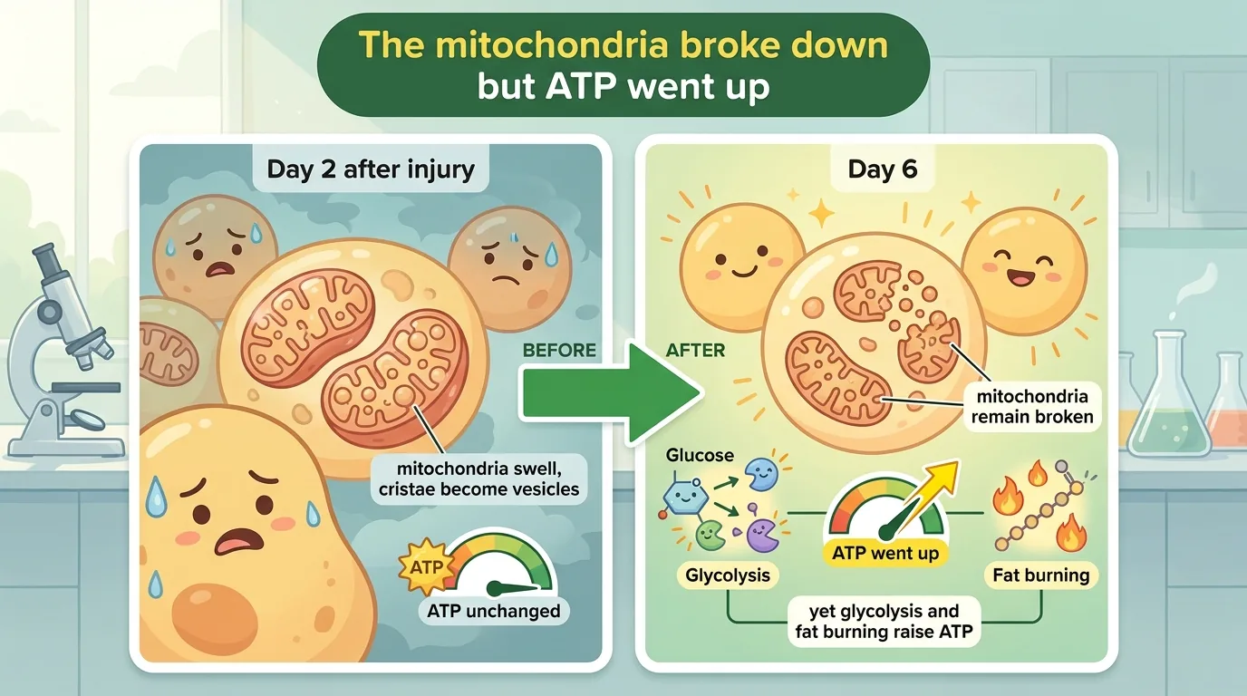
What the Chilean research team looked into this time was the cell’s own “power plant” — the mitochondrion. And there they ran into a scene that defied expectation. In the spinal cord of the frog that cannot regenerate, mitochondria were indeed broken by day 6 after injury. They had swollen, the inner membrane had curled up into vesicle-like shapes, and the enzymes of respiration had stopped working. And yet ATP, the currency of energy, did not fall — it rose.
A wrecked power plant, and more energy. What was going on behind this seemingly impossible combination? And was it really the cells balancing the books? Let us work through it in order.
Publication information
- Paper title: Spinal cord injury induces mitochondrial dysfunction and metabolic reprogramming in non-regenerative Xenopus laevis
- Authors: Miguel E. Domínguez-Romero, Maximiliano Villarreal, Camila Cordero-Véliz, Sebastián Quijada, Francisco Aguirre, Caterina Hernandez, Sol Torruella-Gonzalez, Johany Peñailillo, Verónica Eisner, Paula G. Slater (corresponding author and lead contact)
- Affiliations: (1) Department of Biological and Chemical Sciences, Faculty of Sciences, Universidad San Sebastián (USS), Los Leones campus, Santiago, Chile; (2) the Laboratory of Neuroregeneration and Metabolism at the Ciencia & Vida Center for Scientific and Technological Excellence of Fundación Ciencia & Vida; (3) Department of Cellular and Molecular Biology, Faculty of Biological Sciences, Pontificia Universidad Católica de Chile
- Journal: iScience (Cell Press / Elsevier), 2026, volume 29, article number 116636 (issue 7; print date July 17)
- DOI / link: 10.1016/j.isci.2026.116636 (indexed as PubMed ID 42491811 / PMC13378342, and the full text is free to read)
- Impact Factor: 4.5 (Journal Citation Reports for the 2025 data year, released June 2026; 4.1 the previous year), reported to place it in the first quartile (Q1) of the Multidisciplinary Sciences category. CiteScore is 7.4 (2025, Scopus). iScience is an interdisciplinary journal launched by Cell Press in 2018, covering everything from life sciences to earth sciences
- Open access: Yes. It has been fully open access (gold OA) since launch, and this paper carries a CC BY-NC 4.0 licence. As long as you credit the source and authors, indicate any changes and link to the licence, you are free to read, copy, translate, and even modify and redistribute it (there is no “ND” no-derivatives clause). However, commercial use requires separate permission
- Review timeline: received December 3, 2025 → revised April 16, 2026 → accepted May 7 → published online June 27 (print issue July 17)
- Ethics approval: approved by the Scientific Ethics Committee (Animal and Environmental) of the Pontificia Universidad Católica de Chile and by the Bioethics and Biosafety Committee of Universidad San Sebastián (protocol numbers 181017006 and 210504022)
- Funding: FONDECYT 11220624, Centro Ciencia & Vida FB210008 (ANID), IBRO RS-3304417299 (all to Dr. Slater), and FONDECYT 1231557 (to Dr. Eisner)
- Conflicts of interest: the authors declare no conflicts of interest
- Use of generative AI: the authors state explicitly that during writing they used ChatGPT solely as an editorial aid to improve the grammar, spelling, syntax, clarity and fluency of the English, that they reviewed and edited the content themselves afterwards, and that they take full responsibility for the published content
- Data availability: the data of this study are said to be available from the corresponding author, Dr. Slater, upon reasonable request (they are not deposited in a public repository). Uncropped western blot membranes are included in the supplementary material
About the corresponding author: The corresponding author of this paper is Dr. Paula G. Slater (registered at her institution as Paula Gabriela Slater Guzmán, ORCID 0000-0003-2601-0613). She is an assistant professor in the Department of Biological and Chemical Sciences, Faculty of Sciences, Universidad San Sebastián (Profesor Asistente, appointed November 2022) and, at the same time, an associate researcher of Ciencia & Vida (Centro Basal), the centre of scientific and technological excellence run jointly by Fundación Ciencia & Vida and the university, where she heads her own Laboratory of Neuroregeneration and Metabolism. She is listed as a collaborating faculty member of the university’s doctoral programme in Cell Biology and Biomedicine.
Her career begins, consistently, at the Pontificia Universidad Católica de Chile. After a bachelor’s degree in biochemistry (2011) and a master’s (2013), she obtained her PhD in 2016, specialising in cellular and molecular biology. Her supervisor was Katia Gysling, and her doctoral topic was something entirely different from her present work: the corticotropin-releasing factor (CRF) system. The two postdoctoral positions that followed built the two pillars of her current research. The first was at Boston College in the United States (2016–2018), where she joined Laura Anne Lowery’s laboratory and studied the cytoskeleton and axon guidance. The second was at the Pontificia Universidad Católica de Chile (2018–2022), where she moved to Juan Larraín’s laboratory and took up the relationship between spinal cord regeneration in Xenopus laevis and mitochondria. The acknowledgement of Larraín’s mentorship in this paper traces back to that period.
She is the first author of the review that treats the very subject of this paper, “Mitochondrial function in spinal cord injury and regeneration” (Cellular and Molecular Life Sciences 2022, volume 79, article number 239), and its list of co-authors overlaps almost entirely with the core members of this iScience paper. The direct predecessor to the present study is the paper examining the regenerating tadpole side (R-stage), “Xenopus laevis neural stem progenitor cells exhibit a transient metabolic shift toward glycolysis during spinal cord regeneration” (Frontiers in Cell and Developmental Biology 2025, volume 13, article number 1529093). There too Slater was both first and corresponding author, so this paper can be described as redrawing the reverse side of the picture she painted on the “regenerating” side, applying the same framing of the question to the “non-regenerating” side.
On the funding side, she was selected as sole principal investigator (Investigador Responsable) of FONDECYT early-career grant 11220624, “Function of mitochondrial transfer in axonal and spinal cord regeneration in Xenopus laevis” (awarded by ANID in 2022) — one of the most exciting topics around right now: the idea that mitochondria are handed from cell to cell. In 2025 she received a Rising Star Award from the International Brain Research Organization (IBRO). Among the 2025 awardees published by IBRO, she is the only researcher based in Chile. She also serves as a board member (treasurer) of the Latin American Society for Developmental Biology (LASDB).
The themes her laboratory pursues are how the cytoskeleton and mitochondria shape axonal development and regeneration, and how metabolism bears on the biological processes at work in the early phase of spinal cord regeneration. In recent years she has also branched out into “mitochondrial transfer” — introducing new mitochondria from outside — into how scaffold properties such as stiffness and electrical characteristics affect regeneration, and into the biomaterials that might serve that purpose. This paper is the work that turns that question toward the side that cannot regenerate.
What was it, exactly, that we did not know?
Spinal cord injury (SCI) is a trauma that severs two-way communication between the central nervous system (CNS) and the periphery; everything below the lesion is paralysed, and motor, autonomic and sensory deficits persist. Its incidence and prevalence keep rising worldwide, and yet there is still no effective treatment.
In mammals, humans included, an environment that forbids regeneration is assembled after injury. Mechanical damage injures axons and triggers cell death nearby, while excitotoxicity and free radical production spread the damage into adjacent territory. A prolonged inflammation then begins, and oligodendrocyte precursor cells, astrocytes and ependymal cells proliferate and differentiate into new astrocytes. Where this ends up is the glial scar. It halts the spread of the injury, and at the same time it obstructs regeneration itself.
👦 Student: The body sabotaging its own regeneration — that seems awfully irrational.
🧬 Dr. Exotaro: It is a firebreak: you fell the trees ahead of the flames to stop a wildfire. The forest will not come back there for a while. A short-term judgement that protects life and a long-term goal of recovering function are colliding in the same place.
And yet in nature there are vertebrates that recover sensory and motor function even after their spinal cord is cut: bony fishes, urodele amphibians, and anuran amphibians during their larval stage. Compare animals that regenerate with animals that do not, and the factors that block regeneration and the factors that promote it come into view. That is the logic of comparative research.
This is where Xenopus laevis earns its keep. Both answers exist within a single species. In the tadpole period (NF 48–52, R-stage) sensory and motor function can be recovered even after the spinal cord is transected, whereas the post-metamorphic NF 66 froglet (NR-stage) has lost regenerative capacity, and its response closely resembles mammalian SCI. At NR-stage, the proliferative response of Sox2-positive neural stem progenitor cells (NSPCs) is slow and weak, the cells differentiate into astrocytes and pile up in the lesion gap, forming a glial scar.
👦 Student: Why go to the trouble of using frogs? Wouldn’t mice do?
🧬 Dr. Exotaro: Because the thing we want to compare is not a difference between species. Between a frog and a mouse, the difference gets lost among species differences; but if only regenerative capacity changes within a single species, then that difference is precisely what points to the conditions for regeneration.
The angle of attack in this paper is mitochondria and metabolism. The polarisation state of the mitochondrial membrane has a large bearing on whether a cell exposed to excitotoxicity and free radicals lives or dies. When proliferation rate goes up, the lead role in ATP production shifts from oxidative phosphorylation (OXPHOS) to glycolysis. There are also reports that, after a stem cell divides, the daughter cell that inherits the old mitochondria and switches to OXPHOS differentiates, while the side with new mitochondria and glycolytic metabolism remains a stem cell.
Some things were already known from this viewpoint. Bulk RNA-seq comparing R-stage and NR-stage (Lee-Liu et al. 2014) and proteomics (from the same group in 2018) showed that the expression of mitochondrial and metabolic pathway components differs between the two. And in 2025 the same author team as this paper (Slater and colleagues) reported that in the regenerating R-stage, NSPC mitochondria are remodelled early and in a coordinated fashion, and a transient metabolic shift toward glycolysis occurs. Mitochondrial dysfunction after SCI has been reported in mammals as well. But how metabolism adapts on the side that does not regenerate was almost entirely unknown.
The question of this paper is this. In the spinal cord of a frog that cannot regenerate, what happens to mitochondria and metabolism after injury?
What did this paper find?
What the authors tracked was the six days following complete transection of the spinal cord. At five time points — uninjured, 6 hpt, 1 dpt, 2 dpt and 6 dpt — they looked at the mitochondria of the ependymal cells around the central canal by electron microscopy, measured protein and transcript levels in spinal cord tissue, stained respiratory function directly on the tissue, and quantified ATP as luminescence emitted by a suspension of dissociated cells. The story starts with “where things sit”, passes through “shape”, arrives at “function”, and ends by defying expectation.
What moved first was “where things sit” and “shape”
Animals were anaesthetised with 0.02% tricaine (MS222); the dorsal part of the sixth vertebra was removed by laminectomy at mid-thoracic level to expose the spinal cord, which was then completely transected with microscissors. Uninjured animals served as controls, and the region examined ran from the fifth to the seventh vertebra along the rostrocaudal axis. hpt stands for hours post-transection (hours after the cut), and dpt for days post-transection (days after the cut).
The protagonists are the ependymal cells that surround the central canal. In immunostaining, cells positive for Sox2 — a marker of neural stem progenitor cells (NSPCs) — form 3 to 4 layers around the central canal, with NeuN-positive cells, marking neurons, spread outside them. The authors magnified this region with transmission electron microscopy (TEM) and divided each cell, starting from the central canal side, into apical (up to just before the nucleus), medial (the nucleus) and basal, i.e. distal (beyond the nucleus).
In intact animals the distribution is skewed: 80% apical, 18% medial, and a mere 2% distal. Ependymal cells beat the many cilia on their apical surface to drive the flow of cerebrospinal fluid in the central canal. The power plant sits next to the site where the electricity is used, as the authors read it. This skew has also been reported in regenerating tadpoles and in other animals.
The first thing that catches the eye is how long nothing moves. At 6 hpt and at 1 dpt, the localisation is essentially unchanged from uninjured. What moves is 2 dpt, when the mitochondria scatter inward: about 58% apical, about 28% medial, about 15% distal. The number per unit of cell cross-sectional area also fell transiently at 2 dpt (p<0.05), and by 6 dpt both the arrangement and the number had returned to baseline. The authors, however, are careful. 2 dpt coincides with the period in which earlier work observed discontinuities or damage in the plasma membrane of ependymal cells, and the drop in number may be partly explained by loss of mitochondria. The authors also raise the possibility that the cilia themselves were lost (a conjecture based on reports from traumatic brain injury).
Next comes shape. The cross-sectional area of a single mitochondrion increased significantly at 2 dpt (p<0.05) and grew larger still by 6 dpt (p<0.01). Mitochondria are not fixed little beans; they change form through fusion and fission.
👦 Student: Fusion, fission — do they really move around that much inside a cell?
🧬 Dr. Exotaro: Think of factory consolidation. Fusion is a rescue measure: merge a damaged factory with a healthy one so they can share parts and blueprints. Fission is the opposite — cut off a section with no prospects and send it for disposal. Both are different answers to the question of what to do with equipment that is starting to fail.
So the authors measured the machinery by western blot. Mfn2, which mediates outer membrane fusion, increased at 2 dpt and returned to baseline by 6 dpt, while OPA1, which mediates inner membrane fusion, showed no significant change at any time point. On the fission side, Fis1 increased at 6 dpt (p<0.01). Tom20, the outer membrane import receptor used as a proxy for mitochondrial mass, also increased at 6 dpt (p<0.05).
Does that mean mitochondria were being mass-produced? The authors themselves question this reading. The RNA-seq that enters here is not data newly generated by this study. It is a reanalysis in which the authors remapped spinal cord RNA-seq published by another group in 2014 onto an updated transcriptome (X. laevis v10.1 from Xenbase). Transcript levels of PGC-1α and TFAM, the two master regulators of mitochondrial biogenesis, show no significant change. Since the control centres are not moving, the increase in Tom20 more likely reflects changes in the outer membrane than biogenesis — that is how the authors read it.
The images at 6 dpt capture a tug-of-war. Mitochondria whose outer membranes are continuous while the inner membranes remain separate — suggesting interrupted inner membrane fusion or fission in progress. There are also images in which a swollen region with sparse cristae and a region retaining clear cristae and matrix granules sit side by side within one and the same organelle.
The fusion machinery dominates in the first half, the fission machinery in the second — the authors call this a biphasic, dynamic regulation.
The interior falls apart — vesicle-like cristae, and mitochondria that cannot respire
The folds of the mitochondrial inner membrane are called cristae. The complexes of the respiratory chain are arrayed along those membrane surfaces. Cristae are the workbenches lined up inside the power plant. The authors sorted the TEM images into three types: orthodox, with clear cristae and a dense matrix; swollen, with an expanded matrix and reduced cristae; and hybrid, an intermediate in which both kinds of region coexist within the same organelle.
In uninjured animals, orthodox accounted for about 80%, swollen about 2%, hybrid about 18%. At 6 hpt and 1 dpt this barely changes, but at 2 dpt orthodox falls to about 35%, with swollen about 40% and hybrid about 25%. At 6 dpt orthodox drops further, to about 20%, with swollen about 35% and hybrid about 45%. And this distribution does not return to normal. In contrast to the skew in localisation and the number, both of which returned to baseline by 6 dpt, only the breakdown of internal structure keeps deepening.
The proportion of mitochondria containing vesicular-like cristae — images in which the folds have become a cluster of small sacs — increased significantly at 2 dpt (p<0.01) and rose further by 6 dpt (p<0.0001). Such images have been reported in hypoxia, oxidative stress and ageing, in mitochondrial disease, and in cells in the late phase of apoptosis after cytochrome c release and loss of membrane potential are complete; experimentally, they also appear upon knockdown of OPA1 and upon suppression of prohibitin or mitofilin. Read against those reports, this can be taken as a sign that the machinery maintaining cristae has been compromised — that is the authors’ interpretation.
When the same group examined the post-injury regenerating tadpole (R-stage), these vesicular-like cristae were not detected. They occur only in the froglet that cannot regenerate (NR-stage) — it is because of this asymmetry that the authors read them as “characteristic of the non-regenerative response”.
👦 Student: If only the shape changed, couldn’t they still work?
🧬 Dr. Exotaro: Remember the workbenches. If the boards curl up, you cannot line the equipment on them. But you cannot declare from photographs alone that they “cannot work”. So the authors added tests that translate shape into function.
First, MitoSOX. Detecting mitochondria-derived superoxide in unfixed sections, they found a significant increase in fluorescence intensity at 6 dpt (Fig S2). But, as the authors themselves note, this is an indicator of stress, not an indicator of function itself.
So they added COX/SDH double-labelling histochemistry. COX (cytochrome c oxidase) is complex IV, and its subunits are encoded by the mitochondrion’s own DNA, so intact mtDNA and correct assembly are required for it to work. SDH (succinate dehydrogenase), on the other hand, is complex II, and because its subunits are encoded by nuclear DNA, it does not depend on the integrity of the mitochondrial genome. If both are healthy the result is brown; if only COX fails, the SDH colour remains and it turns blue-purple.
In uninjured animals the cells adjacent to the central canal are uniformly brown, and at 6 hpt, 1 dpt and 2 dpt there is essentially no change. What changes is 6 dpt, when numerous blue-purple cells appear in and around the lesion. Most striking are the lesion core and the region caudal to the injury. Following serial sections along the dorsoventral axis from +50 μm dorsal to −150 μm ventral, the authors found that the COX deficiency was not confined to a single section but extended across multiple depths, radiating several hundred μm along both the rostrocaudal and dorsoventral axes from the epicentre.
The authors’ summary runs as follows. Mitochondrial dysfunction begins to arise around 2 dpt and grows through 6 dpt. It overlaps with the time course over which vesicular-like cristae accumulate.
Broken, and yet ATP had gone up
The authors dissociated the caudal spinal cord fragment with papain, normalised to 80,000 cells per well, and measured luminescence with CellTiter-Glo 2.0. Oligomycin (an inhibitor of ATP synthase) served as a control to gauge the contribution of mitochondrially derived ATP.
From 6 hpt to 2 dpt, ATP does not change. This is consistent with the period in which COX and MitoSOX had not yet moved and respiratory function was relatively preserved (shape had already begun to break down at 2 dpt). But at 6 dpt — the very point at which area was maximal, the hybrid type most abundant, and COX deficient — ATP increased significantly (p<0.05).
👦 Student: It’s broken, and yet the energy goes up?
🧬 Dr. Exotaro: The main power plant has shut down and the emergency generator (glycolysis) is running — that is one way to read it. It is also possible they have started dipping into stored fuel (fat), but there we have only seen that the genes for hauling fuel in are up; nobody measured the site where it is actually burned. And the meter may be picking up electricity that has leaked outside as well.
The authors set out two interpretations side by side. The compensation account: glycolysis and fatty acid β-oxidation (FAO) are mobilised, and energy homeostasis is maintained even under mitochondrial damage. The pathology account: ATP that has leaked out of cells along with tissue damage and inflammatory signalling is simply accumulating. They also state explicitly that the ATP assay used here cannot distinguish the intracellular pool from the extracellular pool, and that therefore both scenarios remain open.
There are three irreversible rate-limiting steps in glycolysis: hexokinase (Hk), phosphofructokinase 1 (Pfk1) and pyruvate kinase (Pkr). Pfkfb is what makes fructose 2,6-bisphosphate, which regulates Pfk1. In newly measured RT-qPCR (with eef1a1 as the internal standard), hk2 rises persistently from 6 hpt and stays high at every subsequent time point. pfkfb1 is unchanged. pklr increases significantly at 6 dpt (p<0.01) — the same time point as the ATP increase.
The authors’ reanalysis of the RNA-seq published by another group in 2014 (no sequence data were generated in this study) points the same way. Glycolytic transcripts increase at 6 dpt, while TCA cycle-related transcripts are essentially unchanged. PDK4, which throttles the entry of pyruvate into mitochondrial oxidation, is also increased. The authors’ conclusion: the enhancement of glycolysis is not coupled to enhanced mitochondrial respiration, and it suggests a shift toward reliance on glycolysis — that is, a partial uncoupling of oxidative metabolism. But also that this shift alone does not fully account for the increase in ATP.
The authors return to the electron microscopy images. At 6 dpt, lipid droplets (LDs) appeared in the ependymal cells adjacent to the central canal and around the lesion — although whether they are fuel is not settled at this point. Gene Ontology (GO) enrichment analysis of the reanalysed data (Metascape) points the same way. What is up is lipid utilisation and transport: “cellular lipid catabolic process”, “lipid homeostasis”, “unsaturated fatty acid metabolic process” and the like. What is down is “cholesterol biosynthetic process”, “sterol biosynthetic process”, “lipid biosynthetic process”. A switch from synthesis to mobilisation and breakdown. On the up side one finds cpt1 (carnitine palmitoyltransferase 1, the rate-limiting enzyme for fatty acid uptake into mitochondria en route to β-oxidation), the nuclear receptor PPARγ, and perilipin (plin); at 6 dpt, ppar-γ and plin2 are increased.
Newly measured RT-qPCR agrees: cpt1 rises mildly at 2 dpt and strongly at 6 dpt (p<0.01). cpt2, which is not rate-limiting, is unchanged at every time point. Oil Red O staining likewise shows nothing in uninjured animals or at early time points, but clear appearance at 6 dpt. It is abundant in cells adjacent to the injury and spreads both rostrally and caudally.
👦 Student: So after sugar comes fat?
🧬 Dr. Exotaro: That is the tempting reading. But β-oxidation is a reaction that uses oxygen inside the mitochondrion. Why would only the genes for hauling fuel into a broken furnace go up?
The answer is the authors’ hypothesis, not a demonstrated fact. All of it is conjecture based on citations. Angiogenesis is said to occur 3 to 7 days after injury, and immature new vessels could partially restore oxygen supply. There are reports that activating FAO with L-carnitine or ketone bodies improved mitochondrial function. And there is the existence of subpopulations: peridroplet mitochondria bound to lipid droplets (said to have high respiratory capacity and ATPase but low β-oxidation) versus cytoplasmic mitochondria. There is no flux measurement whatsoever, and both the rise in cpt1 and the constancy of cpt2 are matters of transcript level.
The following are alternative interpretations the authors raise as citations, and were not observed in this study’s frogs. Lipid droplets increase under stress, in ageing, and in starvation alike. In yeast, contact between the two increases and is said to act as a detoxification mechanism, shunting toxic lipids and proteins into lipid droplets and extending lifespan. In mammalian SCI, large amounts of myelin-derived lipid debris are reported to be generated, and the macrophages that engulf it become foamy macrophages. Plin2 impedes fatty acid mobilisation and protects lipid droplets from degradation, and their formation is also induced by PPAR-γ. Rather than having increased in order to be burned, they may be the result of increased formation and decreased breakdown.
Whether lipid droplet formation after SCI mainly supports energy metabolism or is a detoxification process, and whether this metabolic switch is required for cell survival, awaits future research. Here the authors lay down their pen.
The side that can regenerate and the side that cannot — what differed, and how
Let me note one thing first. What follows about “the side that can regenerate” is not an experimental result of this study. It is what the same group reported in their earlier paper (Front. Cell Dev. Biol. 2025), which this paper cites as the comparison partner.
According to that earlier paper, on the side that can regenerate — the NF 48–52 tadpole (R-stage) — the response is fast, and coordinated. Within 24 hours of transection, transcripts involved in mitochondrial fission were said to rise transiently. Morphology tilts from orthodox toward swollen, but it is like a storm passing through: it returns to its original state by 2 dpt. The mitochondria do uncouple, but overall function is maintained. Metabolism, too, merely shifts transiently toward glycolysis. And the vesicular-like cristae found here in the non-regenerative stage were reported not to be detected in the regenerative stage.
The NF 66 froglet (NR-stage), by contrast, is slow, biphasic, and does not go back. The authors read this difference as the consequence of a mitochondrial adaptive response that is delayed and inefficient. Because the response is delayed, damage to the mitochondria near the lesion becomes greater, forcing a more sustained and larger-scale remodelling that mobilises both the fusion and the fission machinery — that is the authors’ conjecture, and this study contains no experiment testing that causal chain. The 6 dpt picture is emblematic. Mfn2, which mediates fusion, has already returned to baseline, and yet mitochondrial area remains enlarged. The authors read this not as evidence that fusion is continuing but as swelling and structural abnormality, that is, a sign of damage. Mitochondrial dynamics have become uncoupled from the recovery of function.
Line it up with other animals and the outline sharpens. Zebrafish restore mitochondrial function and regenerate. Mice and rats do not restore it, and it worsens tissue damage and obstructs regeneration. The pattern shown by NR-stage Xenopus laevis clearly resembles the mammalian side. That is why this experimental system matters for thinking about human spinal cord injury.
What is it that “the side that can regenerate has and the side that cannot lacks”? The candidate answer this paper offers to that question is speed, and the power to return to baseline. But everything laid out so far is correlation, not causation.
How might the future change? (The road to the clinic)
Let me be blunt first. This study did not create a treatment for spinal cord injury. What was examined is the African clawed frog, and what is being looked at is the shape of an organelle inside cells and the amounts of the molecules tied to it. Whether the ability to regenerate changes when metabolism is blocked or boosted with drugs — that was never once tested in this paper.
With that said, there are three lines of thinking that could connect to the clinic. Please take all of them as possibilities, not conclusions.
The first is the time axis as a blueprint. In the spinal cord that cannot regenerate, mitochondrial abnormality began at 2 dpt and appeared clearly as dysfunction at 6 dpt. Not everything is decided at the instant of injury; the collapse takes several days. If a similar delay exists in humans, then there is a window of time in which to intervene. It could lead to a shift in thinking, from “once the acute phase is over it is too late” to “there are still a few days in which we can act”.
The second is the idea of making metabolism itself a therapeutic target. Among the prior work the authors cite are reports that boosting fatty acid β-oxidation with L-carnitine or ketone bodies improved mitochondrial function and advanced tissue recovery, and an older report that 2,4-dinitrophenol, a mitochondrial uncoupler, improved mitochondrial function in a spinal cord contusion model, reduced oxidative damage and spared white matter. But to be emphatic: this paper tested none of these agents. What this study offers is not a drug but a map for designing “at what moment, and at what to aim”.
The third is a reading of these findings as biomarkers. COX/SDH double staining paints the decline of respiratory function as colour directly on the tissue. And that dysfunction did not stop at a single section: it extended several hundred μm along both the rostrocaudal and the dorsoventral axes. A region wider than the visible lesion is already metabolically damaged. It suggests that in the clinic, too, the extent visible on imaging may not be the extent of the damage.
👦 Student: So if you set metabolism right, does it start regenerating?
🧬 Dr. Exotaro: That is exactly where we must be most careful. Whether the metabolic rewiring is the cause of the failure to regenerate, merely a consequence of the dysfunction, or an ally keeping injured cells alive — that is not settled. And if it turned out to be an ally, an intervention that blocks it with a drug would amount to stepping on the hose while the fire is being put out.
So what is needed next is measurement, not conjecture. Directly measuring how much glycolysis and fatty acid β-oxidation are actually running. And actually pressing or releasing the metabolic switch to see how cell survival and tissue damage move. What we have here is not a prescription but a blueprint for the questions. From here on, let us look squarely at the weak points of that blueprint.
How to read this study critically — limitations, and routes to higher quality
Start with what deserves credit. First, the statistical design. Because TEM measures many mitochondria from a single animal, the data have a nested structure. Applying nested one-way ANOVA here is appropriate; it avoids inflating the number of measurements within an animal into independent n. Second, the way the readouts are assembled. What MitoSOX sees is oxidative stress, not function itself — the authors judged as much and added COX/SDH double labelling. Contrasting mtDNA-encoded complex IV with nuclear-encoded complex II on the same section is a clever design. Third, they systematically traced the spread of COX deficiency along both the rostrocaudal and the dorsoventral axes. Fourth, the very choice of a model in which a regenerating and a non-regenerating period can be compared within the same species is a strength. Fifth, they list the limitations frankly, and for the ATP increase at 6 dpt they do not adopt the compensation reading alone but set the pathology-side explanation, accumulation of extracellular ATP, alongside it. Sixth, their handling of transparency is honest, down to including uncropped western blot membranes and disclosing generative AI use and conflicts of interest.
There are limitations the authors themselves state explicitly. The metabolic changes are inferences drawn from morphology, ATP, transcripts and histochemistry; no direct metabolic flux measurement was performed. Many of the analyses were carried out on whole spinal cord tissue (homogenate), so metabolic responses cannot be distinguished by cell type. There are no functional experiments directly testing whether mitochondrial dysfunction and metabolic reprogramming contribute to the failure of regeneration or to cell survival — it is correlation, not causation. The ATP assay cannot distinguish the intracellular pool from the extracellular pool. NF 66 animals are not yet sexually mature and their sex cannot be determined, so no analysis of sex differences is possible.
There are points to add from the reader’s side. The n is small — at least 3 independent matings, with 3 to 4 animals per time point — and there is no statement of a power calculation (sample size is stated to be based on previous work and experience). While they say quantification was done on coded specimens wherever possible, they also write that complete blinding was not always feasible for surgery and tissue processing. Since the orthodox / swollen / hybrid morphological classification involves observer judgement, that is where incomplete blinding bites hardest. The RNA-seq is a reanalysis of data published by another group in 2014, derived from different animals and different experiments than the TEM and ATP of this study, so the alignment of time courses is indirect. The GO analysis specifies human (Homo sapiens) for both input and output, so information on frog-specific genes and on .L / .S homeologs may be lost. Transcript levels (mRNA) are not enzyme activity, and there is no guarantee that a rise in cpt1 means enhanced β-oxidation. The data are not in a public repository and are available only on “reasonable request” to the corresponding author. The time points also stop at 6 dpt, so we do not know whether this metabolic state persists into the chronic phase or resolves.
So how could the quality be raised? First, directly quantify glycolytic rate and fatty acid oxidation rate separately, using extracellular flux analysis such as Seahorse or 13C-labelled tracers. Second, raise the cell-type resolution — isolate ependymal cells by FACS, or overlay metabolic imaging on single-cell or spatial transcriptomics. Third, interventions that ask about causation. When the metabolic switch is blocked — glycolysis with 2-DG, CPT1 with etomoxir, or PPARγ inhibition — how do cell survival, tissue damage, and limited regenerative indices move? Conversely, does promoting FAO with L-carnitine improve things? Add to that: measuring ATP in the perfusate and separating intracellular from extracellular ATP by apyrase treatment; generating new RNA-seq from this study’s own animals so that the time course aligns with the TEM and ATP; full blinding of the morphological classification and reporting of inter-rater agreement (κ); direct measurement of membrane potential with TMRM and of oxygen consumption; and extension into the chronic phase over weeks to months.
Even with all of that laid out, the value of this study does not fall. No one had described, with coordinates in both time and space, the order in which the position, number, shape and function of mitochondria break down in the spinal cord of a frog during its non-regenerating period, and which way metabolism swings. Causation is untouched, and this is preclinical work — basic research in an amphibian at that. But as a map pointing to what should be measured next, it is more than sufficient.
Dr. Exotaro’s Perspective
I myself have studied the treatment of spinal cord injury (SCI) with mesenchymal stem cells (MSCs) and their extracellular vesicles (EVs). So this paper felt to me like something sitting squarely in my own arena while striking the same wall from a completely different angle. While we are thinking about what to deliver, this paper is peering through an electron microscope at what the receiving cells are losing at that moment. Let me note first that neither exosomes nor extracellular vesicles appear even once in this paper. Everything I write below, drawing it toward my own speciality, is my personal opinion.
What struck me first was this one point: “does not regenerate” did not mean “does nothing”. The spinal cord of the frog in its non-regenerating period rearranged the placement and shape of its mitochondria, belatedly but nonetheless, raised in concert the transcripts involved in glycolysis and in fat mobilisation, and by 6 dpt had if anything more ATP. Here the paper itself is careful, declining to decide whether this is compensatory metabolic reprogramming or an accumulation of extracellular ATP leaking from damaged tissue. Even so, it looks less like quiet resignation than like a desperate balancing of the books — though whether the books actually balance is, as we saw, unsettled. And precisely for that reason, as someone thinking about treatment, I want to ask myself again whether my own intervention is getting in the way of that balancing act.
The other point is speed, and the power to return to baseline. What separated R-stage from NR-stage was not whether change occurred but how quickly the change got going and how far it could return to the original state. I think what the clinic calls the “therapeutic window” might be recast not in terms of the names of molecules but as a metabolic time axis.
From here on it is my imagination. When MSC-derived EVs work in spinal cord injury, the way they work has been described in terms of suppressing inflammation or protecting axons — but the possibility that they are rebuilding the energy state of the recipient cells themselves is something I have long wondered about. In recent years there have also been reports that mitochondria themselves, or fragments of them, are handed between cells. To repeat, this paper did not test that. Still, the concrete picture of ependymal cells losing the ability to respire as they head toward 6 dpt gives us a clue for thinking about what we should be delivering.
Let me put it back in place at the end. This is preclinical basic research using an amphibian, and it does not connect directly to human spinal cord injury. Even so, I think it is a study that lifted a corner of the curtain and showed us what is happening on the other side of the wall.