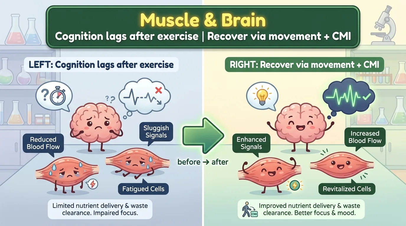
“Exercise is good for the body”—everyone knows that. But what if you were told “exercise is good for the mind” too? The idea that aerobic exercise, such as walking or cycling, can also lift cognitive functions like memory and attention has spread rapidly over the past decade. Yet in the actual clinic of neurological rehabilitation, something else often happens: “the good markers in the blood went up after exercise, but the patient’s cognition barely changed.” What I am introducing today is a review that confronts the true nature of that discrepancy head-on. The key turned out to be an unfamiliar concept: cognitive-motor integration (CMI).
Journal Information
- Paper title: Muscle and mind: rewiring cognitive-motor recovery through exercise-responsive neurophysiology in neurological populations
- Authors: Andrea Calderone, Alessio Baricich, Sanaz Pournajaf, Fabrizio Sottile, Maria Grazia Maggio, Mirjam Bonanno, Rosaria De Luca, Francesco Ligato, Angelo Quartarone, Rocco Salvatore Calabrò (IRCCS Centro Neurolesi Bonino Pulejo 〈Messina〉 and others, Italy)
- Journal: Frontiers in Psychology (2026, Vol. 17, Article 1845070, published online June 17, 2026)
- DOI / link: 10.3389/fpsyg.2026.1845070
- Impact Factor: roughly around 2.6 (an open-access journal broadly covering psychology and neuroscience)
- Open access: yes (CC BY. Free to reuse with proper attribution). The type is a “narrative review”—rather than reporting a single experiment, it re-stitches into one line of reasoning the evidence scattered across the separate traditions of exercise physiology, neurorehabilitation, movement science, and cognitive medicine. The literature was gathered from PubMed/MEDLINE, PsycINFO, Embase, and Scopus as of March 16, 2026.
- Transparency note: the authors state explicitly that they used generative AI (Gemini 3.1 Pro) only in a limited way for creating Figures 1–4 and for technical editing, that they used none of it for generating the text, citations, or arguments, and that everything was verified and approved by the authors.
What kind of researcher is the corresponding author? The first and corresponding author, Andrea Calderone, is an early-career researcher in neurorehabilitation who lists as this paper’s affiliation the neurorehabilitation research hub IRCCS Centro Neurolesi Bonino Pulejo in Messina, Sicily, Italy (public sources also indicate ties to the University of Messina). On themes such as cognitive rehabilitation, rehabilitation robotics, virtual reality (VR), tele-rehabilitation, the digitalization of neuropsychological testing, and the use of AI in neurological disease, this author has been energetically publishing numerous papers, including systematic reviews. The senior author, Rocco Salvatore Calabrò, is a high-output researcher who has led neurorehabilitation research at the same facility for many years, and co-author Angelo Quartarone serves as the scientific director of that IRCCS. It can be positioned as a review in which one of Italy’s foremost neurorehabilitation research teams has organized a vast body of knowledge on “exercise and the brain” into a single map.
What, exactly, was previously unknown?
Neurological rehabilitation has long treated the “recovery of motor function” and the “recovery of cognitive function” as separate tasks. Physical therapy handles gait and the movement of the limbs; occupational therapy and neuropsychology handle attention and memory—that division of labor is easy to understand as a clinical convenience. But our daily life is not built that way. Walking down a crowded corridor while carrying a bag, dodging people, answering when spoken to, and preparing to reach for a doorknob—this chain of actions mobilizes postural control, monitoring of the surroundings, movement planning, and executive function (EF) simultaneously. Walking is by no means “a job for the legs alone.”
The concept this review places at its center is cognitive-motor integration (CMI). CMI is defined as the ability to mesh “cognitive control” with “motor execution” so that purposeful movement remains accurate, flexible, and situationally adaptive even as the environment or one’s own state changes. There is a similar term, “dual-task,” but CMI is broader—it refers to the supple coupling of cognition and movement itself.
👦 Student: Exotaro, are cognition and movement really that inseparable?
🧬 Exotaro: In everyday life, yes. While you’re healthy, you never even notice that you “think while walking.” But in stroke or Parkinson’s disease, walking itself uses up your attention. So the moment you start thinking, your feet stop; or the moment you move your feet, the conversation stops. Cognition and movement compete for resources. How to untangle that competition is the heart of rehabilitation.
And now the main issue: the discrepancy. Much of the story that exercise is good for the brain rests on the observation that exercise raises “seemingly good markers” in the blood, such as BDNF (brain-derived neurotrophic factor). Yet when it comes to whether patients with neurological disease actually improve cognitively, the effect is often “so-so” and “variable.” The marker moved, but cognition in daily life did not return as much as hoped. Why?
The authors’ view is simple: “a biomarker changing” and “cognition recovering” are different matters. Exercise may prepare the groundwork for the brain to change, but whether that becomes a genuinely useful change depends on whether that biological state of readiness is actually used within meaningful, attention-demanding movement. This review draws a three-step line of reasoning—(1) through what peripheral signals does exercise reach the brain, (2) how is it “translated” within the brain, and finally (3) why does it acquire clinical meaning only once it surfaces as the “behavior” of CMI—organized around four diseases: stroke, Parkinson’s disease, multiple sclerosis, and traumatic brain injury.
What did this paper find?
Exercise “sends letters from muscle to brain”—the peripheral mediators
The starting point is the recognition that “muscle is not merely an organ that receives commands and contracts.” Contracting muscle releases signaling substances called myokines and metabolic messengers, acting as a “signal-emitting organ” that alters the tone of inflammation and the state of the vascular, immune, and endocrine systems. When exercise works on the brain, its effect is delivered mainly through such peripheral messengers. The review organizes the representative messengers.
BDNF is the most-discussed protagonist. It supports the maintenance of synapses, the remodeling of dendrites, and long-term potentiation (LTP)—the cellular mechanism of learning—thereby promoting activity-dependent learning. Its blood concentration can rise with exercise. But the authors are level-headed: because BDNF in the blood also comes from outside the brain—platelets, vascular endothelium, immune cells—its tissue specificity is low, and it cannot be treated as “direct evidence that the brain was repaired.” BDNF can serve as an indicator that “a plasticity-related system was engaged,” but nothing more and nothing less.
The FNDC5–irisin axis likewise symbolizes both hope and uncertainty. When exercise moves the signaling of PGC-1α (peroxisome proliferator-activated receptor-γ coactivator 1α)/FNDC5, adaptations related to BDNF expression and memory in the hippocampus can be promoted. However, interpretation in humans is limited by measurement methods and tissue specificity, so it should be presented not as a “clinically established biomarker” but as a “biologically plausible intermediary.”
IGF-1 (insulin-like growth factor 1) is another endocrine route. It changes with exercise, crosses the blood-brain barrier (BBB), coordinates with BDNF-related signaling, and is involved in synaptic support and metabolic adaptation. Here, too, the cautious reading recommended is not “high IGF-1 = recovery,” but a permission signal that opens “a biological window in which learning is more likely to occur.”
And the one undergoing reappraisal is lactate. It used to be spoken of through the popular image of “a fatigue substance that accumulates during exercise,” but the original paper re-frames it more neutrally as “merely a byproduct of anaerobic metabolism.” Lactate is now recognized as another fuel for the brain and re-conceived as a “signaling molecule” that crosses the BBB via monocarboxylate transporters and interacts with SIRT1-dependent pathways and BDNF-related signaling. Lactate can serve as a marker that “a metabolically meaningful load was applied”—but “more is better” does not hold. In neurological disease, because of fatigue, autonomic dysregulation, and heat sensitivity, even a low exercise intensity can produce large internal stress. The lactate shuttle hypothesis shows that lactate also connects, via the HCAR1 receptor, to the brain’s VEGF (vascular endothelial growth factor) and angiogenesis pathways, adding still more dimension to the picture.
The immune, vascular, and endocrine “environment” cannot be overlooked either. With exercise, muscle releases IL-6 (interleukin-6) as a myokine, which can promote the anti-inflammatory IL-10 and the IL-1 receptor antagonist, working to quiet chronic low-grade inflammation. On the vascular side, endothelium-derived nitric oxide signaling supports cerebral blood flow. Because cognition depends on “blood flow reaching the right place at the right time,” this underpinning of blood flow is important.
👦 Student: If so many messengers help the brain, then the more you exercise, the better cognition should get……
🧬 Exotaro: That’s exactly the trap. The authors do not call these messengers “the direct cause of recovery.” They describe them strictly as an internal biological dose—the groundwork that raises or lowers the probability that the brain can learn. The letter was delivered. But if that letter isn’t read and acted upon, no meaning arises. This is the single most important point today.
How is it “translated” inside the brain?—the central conversion
Peripheral signals acquire meaning only once they are translated into central changes that alter cognition and CMI within the brain. The central mechanisms the review lists are synaptic plasticity, cellular energy metabolism, signaling related to neurogenesis, neurovascular coordination, and network efficiency.
Synaptic plasticity and LTP are the core of the theory linking exercise and cognition. In humans, too, studies using transcranial magnetic stimulation (TMS) have shown that a single bout of aerobic exercise can enhance LTP-like plasticity in the motor cortex. This is a measurable bridge showing that “exercise temporarily prepares a state of readiness for plasticity.” But the authors draw a line: this itself does not prove cognitive recovery; it is merely evidence that “learnability went up.” Moreover, this “window of learnability” is meaningless unless it is used at the right behavioral timing—for stroke, visuospatial relearning; for Parkinson’s, cue-dependent gait control—so the task to be combined differs by disease.
Signaling related to neurogenesis (especially in the hippocampus), neurovascular coupling (the mechanism that delivers blood flow in step with local neural activity), and the reorganization of large-scale networks are also organized as pathways by which exercise can support cognition. But what they share is the same admonition: even when a biomarker moves, a measurable improvement in cognition may not follow. If the time window in which biology moves and the time window in which behavior is tested are out of phase, the effect is invisible. Conversely, even without a clear change in peripheral markers, function can improve in terms of task learning, strategy use, and improved real-world tolerance (ecological tolerance). This misalignment is not a failure but information, the authors say—it is evidence that central translation is strongly shaped by the location of the lesion, the disease stage, the medication state, task selection, and the behavioral context.
Cognitive-motor integration (CMI)—it acquires meaning only when it surfaces as “behavior”
This is the backbone of the review. CMI is the behavioral-level interface where biological readiness rises as function. A patient responding to information while walking, dodging an obstacle, reaching in time, correcting an error, judging whether to stop—these behaviors mobilize executive function, sustained attention, working memory (WM), sensory prediction, and fine motor adjustment simultaneously. Monotonous aerobic exercise can improve vascular and systemic physiology, but recovery specific to CMI requires that that prepared internal state actually be summoned within tasks demanding “coordination, interference management, prediction, inhibition, and updating.”
👦 Student: So rehabilitation where you use your head while walking is better than rehabilitation where you just walk?
🧬 Exotaro: As a direction, yes. Dual-task training strengthens the cognitive-motor demand and tests “whether the prepared biology is being translated into interference management.” But the authors are cautious: with CMI, calibration is everything. To expose the bottleneck it has to be hard, but it must not be so hard that it wrecks the safety or quality of the movement. Imposing an unreasonable dual task on someone with an unstable gait only raises the risk of falling. Set the difficulty where it is “safe yet challenging”—that is the core of the prescription.
Another pillar is error-based adaptation and sensorimotor prediction. The quality of movement hinges on predicting the outcome, detecting the deviation, and stabilizing the next move. This computation is vulnerable to fatigue, distraction, and disease-related processing delays. That is precisely why, even in a biologically active exercise session, if the practice that follows is too passive, too simple, or divorced from decision-making, it will not lead to meaningful recovery—CMI tasks should be chosen not to “add difficulty” but to target “the cognitive operation that is the patient’s limiting factor,” it is emphasized.
The “manner of translation” differs by disease
Even with the same external prescription of “exercise,” the internal dose it produces and how it works on cognitive-motor function change greatly by disease. The review carefully depicts these differences across four neurological diseases.
In stroke, the location and size of the lesion, the time since onset, vascular reserve, hemispatial neglect, aphasia, and prior cognitive burden govern the internal dose of exercise. A protocol that works in one subgroup does not work in another—because the limiting factor differs: is it insufficient perfusion, reduced endurance, unstable attention, or inefficient relearning? If there is visuospatial neglect, use gait and reaching tasks that scan the environment and dodge obstacles; if executive function has declined, use graded response selection and set-switching during walking—that is, tasks are assembled to match the function that is impaired.
Parkinson’s disease (PD) is a distinctive case in which declines in executive function, set-switching, visuospatial processing, and automaticity are entangled from early on. Exercise can act on dopamine-sensitive circuits, frontostriatal efficiency, gait automaticity, and responsiveness to cues. But clinical results are “promising but not uniform.” Moreover, in the “on” state, where medication is working and compensatory control is possible, effects look large, while in states dominated by freezing of gait, fluctuations, postural instability, and cognitive fatigue, they look small. Rhythmic auditory cues, visual footfall targets, and turning tasks with response inhibition train the ability to “compensate with attention” when automaticity has broken down. The same cognitive challenge improves cue use in one patient but destabilizes freezing or posture in another—this individual variability is precisely what makes designing CMI in PD difficult.
Multiple sclerosis (MS) is a context dominated by inflammation, demyelination, fatigue, heat sensitivity, and fluctuating disability. Exercise can improve deconditioning and mood and act on inflammation and network efficiency, but the response is conditioned by disability level, fatigue, and relapse status. Here CMI should be built around not “maximum difficulty” but “fatigue-conscious processing speed,” and the conservative framing recommended is to treat next-day symptom worsening not as a “compliance problem” but as a “dose signal.”
Traumatic brain injury (TBI) makes the translation problem even harder, because injury severity, symptom profile, sleep, autonomic stability, vestibulo-ocular symptoms, and exercise tolerance vary greatly from patient to patient. Exercise is biologically reasonable when seen through the frame of cerebral perfusion, mood, autonomic recalibration, sleep, and graded return to activity, but while headache, dizziness, sleep disturbance, and visuomotor hypersensitivity persist, an ordinary aerobic prescription can turn into “symptom-provoking exposure.” So CMI requires a graded design: start below the symptom threshold and only advance once symptoms have returned to baseline.
How will the future change? (the road to the clinic)
Use technology “to match the purpose”
VR, robots, wearables, and non-invasive brain stimulation (NIBS)—these technologies, depending on how they are used, strengthen the CMI framework. VR can manipulate task complexity, salience, reward, and contextual variability; robots raise the precision of repetition, assistance, resistance, timing, and measurement; wearables capture gait variability, fall risk, and symptom fluctuation outside the clinic; and NIBS can tilt plasticity in a targeted direction. But the authors’ consistent argument is: “technology is not decoration—choose it to match the translation problem you must solve.” As in the example where robot-assisted hand therapy after stroke can precisely combine somatosensory and cognitive demands rather than mere repetition, value emerges only from mechanism-matched use.
Prescribe by “internal load,” not “external workload”
If exercise is to be used as “a tool that works on cognition,” the prescription must design modality, intensity, duration, sequencing, and task context to match the biological pathway and the behavioral demand. Here the authors urge us to re-conceive intensity not as “external wattage or speed” but as “internal biological load.” Even at the same treadmill speed, the internal meaning differs entirely from patient to patient. Individualization arises as “four linked judgments”—looking all the way at the disease context, baseline fitness, fatigue, medication state, autonomic stability, and tolerance to cognitive load: (1) decide the tolerable biological exposure, (2) choose a clinically meaningful movement, (3) choose the cognitive demand that mirrors that patient’s limit, and (4) monitor whether the combined task maintains its quality.
👦 Student: So in the end, there’s no all-purpose exercise where “if you do this, cognition recovers”?
🧬 Exotaro: That’s right. The core of this review comes down to one line: “exercise is not a panacea but a targeted exposure.” The same prescription becomes a vascular stimulus for one person, a fatigue-inducing load for another, and a meaningful CMI task for yet another. Only once you specify all of this—“for whom, under which disease constraints, at what internal dose, through which pathway, combined with what behavioral task, toward what real-world outcome”—does exercise become cognitive rehabilitation.
Design the next generation of trials by “alignment”
The design principle the review raises repeatedly is alignment. Match the exercise dose to the pathway markers, match those markers to the central readout, match the central readout to the CMI task, and match the CMI task to the ecological (real-world) outcome. Only this chain can judge whether “exercise-responsive biology became clinically meaningful cognitive-motor recovery”—and, they argue, it should be specified in advance, not narrated after the fact once the results are in. Only in this way does the review’s integrative model turn into “a rehabilitation framework that can be experimentally refuted.”
Regarding new biomarkers, too, the authors call not for a “list” but for “alignment with pathways.” VEGF (an indicator of vasculature and perfusion), cathepsin B (a memory-related signal induced by running), the kynurenine pathway (a route of neuroactive metabolites from muscle to brain), GPLD1 (a liver-derived exercise-related mediator), and β-hydroxybutyrate (a ketone body involved in BDNF transcription)—each acquires meaning only when measured after predefining the sampling time window, the tissue source, the central readout, and the behavioral endpoint. The stance is to turn candidate markers into “testable hypotheses.”
In short—the shortest path “from muscle to mind” is not reductionism. It is being integrative, disease-sensitive, and behaviorally precise. Biomarkers tell us about the process, but they cannot substitute for behavior. The decisive step is not proving that “exercise alone changed molecules, perfusion, or networks,” but proving that “a disease-appropriate biological exposure was coupled with a disease-appropriate CMI task and expressed as safer, more flexible, more durable behavior.”
How should we read this study critically?—limitations, and paths to raise its quality further
A good reader never swallows things whole; they always ask “how certain is this?” As Exotaro, I apply the same measuring stick to this review as well.
Limitation ①: this is a “narrative review,” not a systematic review. The authors deliberately chose this format because the question is integrative, and they follow the conventions of a transparent narrative review (Gasparyan et al., 2011, and others). But the price is that there is no pre-registered protocol (such as PROSPERO), no PRISMA flow diagram, no report of the number of selected references, and no risk-of-bias assessment for each study. The databases searched (PubMed/MEDLINE, PsycINFO, Embase, Scopus) and the update date (March 16, 2026) are shown, but inclusion and exclusion are a selection by “relevance” and “priority,” so which studies were included and excluded cannot be strictly reproduced, and the possibility that counter-evidence was underweighted cannot be fully ruled out. → What could have been done better: making it a systematic (or scoping) review with a pre-registered protocol + explicit selection criteria + PRISMA + a GRADE certainty assessment would make the evidence foundation auditable. Furthermore, for the effectiveness question “is dual-task/CMI training superior to usual care,” a meta-analysis computing effect sizes by disease could have replaced “so-so but meaningful” with numbers.
Limitation ②: the central model is an “attractive hypothesis” that has not yet been tested. The integrative model (Figure 1) and the “alignment” principle are conceptually beautiful, and the authors themselves candidly admit they “should be pre-specified” and “should be made falsifiable.” But this review itself does not test it—it is a proposed framework, not a confirmed hypothesis. Much of the mechanistic chain is supported by “may” and “could,” often extrapolating animal and preclinical findings to human patients. → What could have been done better: running the framework on at least one worked example. Re-analyzing an existing clinical trial to show that the chain “a specific exercise dose → a biomarker with a defined sampling window → a central readout (e.g., TMS or perfusion) → a disease-matched CMI task → a real-world outcome” can actually be measured would turn the figure into “a testable protocol.”
Limitation ③: breadth at the expense of depth. It packs 4 diseases × many mediators × many technologies into a single paper, so you get a map but the measured values at each node are scarce. (To be fair, Table 2 presents human evidence and preclinical evidence separately, and this honesty deserves credit.) → What could have been done better: combining the narrative map with a quantitative appendix that adds disease-specific effect sizes, heterogeneity, and a data-backed decision tree for the “four linked judgments” would give clinicians not just “principles” but “a calibrated guide.”
Limitation ④: a note on independence. The disclosure posture is honest—but the authors were editorial board members of Frontiers at the time of submission, and the handling editor declared prior co-authorship with some of the authors. This does not invalidate the conclusions, but it is only right to read the claim “our framework is the way forward” with healthy skepticism. The use of generative AI (Gemini 3.1 Pro) is also disclosed as limited to the creation and editing of figures, and the scope is appropriately restrained.
All in all—none of this sinks the paper. A narrative review is the best tool for sewing scattered fields into a single logic, and the claim that “biomarker ≠ behavior” is, if anything, valuable in that it guards itself against overreaching assertions. But the honest assessment is this: this is a persuasive “hypothesis-generating map,” not evidence in itself that the map is correct. Its true worth will be tested from here on by the next generation of trials that this review calls for.
Exotaro’s perspective
To be honest, this paper is not an exosome paper. The extracellular vesicles (EVs) and mesenchymal stem cells (MSCs) I usually deal with barely appear. And yet I took up this review because the skeleton of thinking written here resonates surprisingly deeply with my own research—how to treat neurological diseases, including spinal cord injury (SCI), using MSCs and EVs.
What I always insist on is not “how much you put in” but “how you deliver it and connect it to function.” No matter how excellent a therapeutic message you load into the natural capsule that is an EV, unless it reaches the target site and is actually used within the injured circuit, the patient’s life does not change. The core of this review—“a biomarker merely moving does not constitute recovery; prepared biology acquires meaning only when it is summoned within meaningful behavior”—was precisely the very question I face in regenerative medicine. Preparing the groundwork for repair with cells or EVs, and translating that groundwork into function. These two are continuous yet separate jobs.
That is exactly why I believe regenerative medicine and rehabilitation are not opposites but a confluence. Even if we raise the repair potential of nerves with EVs, that repaired tissue, in the end, must be trained within “attention-demanding movement” and rise up as the behavior of CMI, or it will not connect to walking or the use of the hands. Conversely, no matter how skillful the rehabilitation, without the biological groundwork for repair it hits a ceiling. Findings are also emerging that exercise itself releases EVs and exchanges information between muscle and brain, and “exercise” and “extracellular vesicles” should eventually connect on a single map.
I was also encouraged that this review came out of one of Italy’s foremost neurorehabilitation research teams. The stance of not being swept up by the glamour of molecules, but calmly re-interrogating all the way to “for whom, under which disease constraints, toward what behavioral outcome,” is the very discipline I want to keep holding in regenerative medicine. Adding just a little of our own wisdom to the natural capsule, and designing it all the way through to “how to deliver it” and “how to translate it into function”—believing that beyond that lies a future where “someone who could not walk stands up again,” I continue my research today.