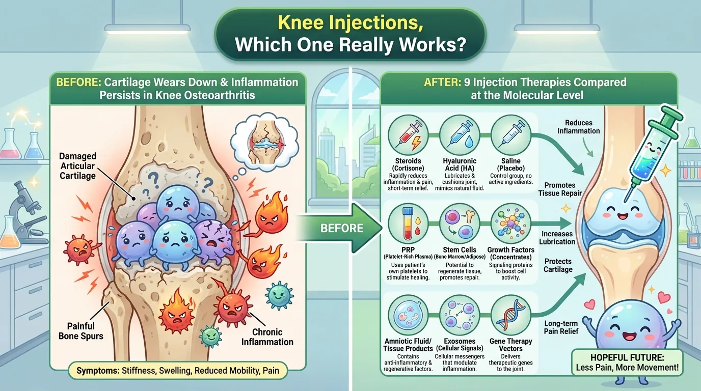
Your knee hurts. You go to an orthopedic clinic, and they ask, “Shall we give you an injection?” But few people realize that there are at least nine different injection options, each working through a different mechanism and backed by wildly different levels of evidence. Today’s paper is a review that surveys intra-articular injection (IAI) for knee osteoarthritis (KOA) at the molecular level—spanning everything from corticosteroids, the “old standby” drug, to exosomes, the “cutting-edge extracellular vesicle” that still remains at the animal-testing stage.
👦 Student: Isn’t an injection just something that kills the pain and that’s it?
🧬 Dr. Exotaro: That’s exactly today’s theme. Even though it’s the same act of “injecting into the joint,” the range runs from things that merely calm inflammation to things that try to protect the cartilage itself. And the “strength of evidence” varies wildly from one treatment to the next. Today, let’s draw that map together.
Journal Information
- Paper title: Molecular Advances in Intra-Articular Injections for Knee Osteoarthritis
- Authors: Juan M. Roman-Belmonte (Department of Physical Medicine and Rehabilitation, Cruz Roja San José y Santa Adela Hospital, Madrid; wrote the initial draft), Hortensia De la Corte-Rodriguez (Department of Physical Medicine and Rehabilitation, La Paz University Hospital-IdiPaz), E. Carlos Rodriguez-Merchan (Department of Orthopedic Surgery, La Paz University Hospital-IdiPaz; corresponding author)
- Journal: Frontiers in Bioscience-Landmark (Front. Biosci. (Landmark Ed)), 2026, Volume 31, Issue 7, 51917
- DOI: https://doi.org/10.31083/FBL51917
- Peer review & publication timeline: Submitted March 16, 2026; revised May 17; accepted May 22; published July 24 (Academic Editor: Elisa Belluzzi)
- Open access: CC BY 4.0 license
- Type of paper: A review paper (review). However, the text contains no mention of the database names used for the search, a PRISMA flowchart, or a registered protocol; methodologically, this is not a “systematic review” but rather a narrative review based on the authors’ knowledge and experience. This point is examined in detail later, in “How to Read This Study Critically.”
- About the journal: Frontiers in Bioscience-Landmark is an academic journal published by IMR Press, based in Singapore. Note that this is a different publisher from the similarly named “Frontiers” series (Frontiers in Pharmacology, Frontiers in Immunology, etc., published by Frontiers Media SA in Lausanne, Switzerland)—be careful not to confuse the two. The NLM Catalog confirms that MEDLINE/PubMed indexes Volume 14 onward, published from 2009. According to the publisher’s official page, it is also indexed in Scopus, SCIE (Web of Science), and DOAJ, which is consistent with the records of several independent indexing sites. As for the Impact Factor, the journal’s official account has announced 3.3, as 2023 Journal Citation Reports data, from Clarivate. Note that the publisher’s website currently also displays a figure of “4.1,” but it does not specify which year’s data this is based on, so it is reasonable to adopt the former figure, 3.3 (2023 JCR data), as the confirmed information.
- Funding & conflicts of interest: The paper states explicitly that “this study received no external funding” and that “the authors declare no conflicts of interest.”
About the corresponding author: E. Carlos Rodriguez-Merchan is affiliated with the Department of Orthopedic Surgery at La Paz University Hospital in Madrid, and with IdiPaz (La Paz University Hospital Institute for Health Research), the hospital’s affiliated medical research institute. This affiliation matches the information listed in another paper by this author registered in PubMed (a sole-authored 2022 review in the International Journal of Molecular Sciences on intra-articular mesenchymal stem cell injection for knee OA, covering its current molecular mechanisms and clinical effectiveness), which was directly verified. In other words, even before writing the present review, this author had already been writing reviews in exactly this field—regenerative-medicine intra-articular injection for knee OA. This author has also continuously worked in the clinical care and research of hemophilic arthropathy, repeatedly publishing papers in the journal Haemophilia together with co-author De la Corte-Rodriguez. Knee joint surgery is likewise one of this author’s specialties, and their name appears as an editor of the Springer book Advances in Orthopedic Surgery of the Knee (2023). Note that no source obtained specified this author’s gender, so this article consistently avoids gender-specific language. Co-author Juan M. Roman-Belmonte, affiliated with the Department of Physical Medicine and Rehabilitation at Cruz Roja San José y Santa Adela Hospital in Madrid, wrote the initial draft of this review; co-author Hortensia De la Corte-Rodriguez, affiliated with the Department of Physical Medicine and Rehabilitation at La Paz University Hospital-IdiPaz, has repeatedly co-authored research on hemophilic joint disease together with Rodriguez-Merchan.
What Wasn’t Understood Until Now?
For a long time, KOA was thought of as a disease of “cartilage wearing away”—a degenerative condition primarily caused by mechanical wear. But this paper emphasizes that this understanding is “incomplete.” KOA is not a disease of cartilage alone; it is a disease of the “whole joint as an organ,” encompassing the cartilage, subchondral bone, synovium, meniscus, joint capsule, and even the infrapatellar fat pad—and, moreover, a persistent, low-grade inflammatory disease involving multiple immune-related signaling pathways.
This inflammation arises from multiple locations throughout the joint. When the meniscus degenerates, gene expression in the surrounding chondrocytes changes in a paracrine manner, inducing cyclooxygenase-2 (COX-2) and matrix metalloproteinase-3 (MMP-3). In the synovium, matrix breakdown products from cartilage and joint tissue stimulate Toll-like receptors and the complement cascade, releasing inflammatory cytokines. The infrapatellar fat pad, alongside neuropeptides, serves as a local source of classic inflammatory cytokines such as IL-1β, IL-6, and TNF. Furthermore, at the level of the nervous system, the paper organizes multiple pathways linked to pain and disease progression—including sensitization of peripheral nociceptors by inflammatory cytokines and nerve growth factor, central sensitization (the brain and spinal cord’s “hypersensitization to pain”), and a phenomenon in which osteoclasts in the subchondral bone produce netrin-1, driving abnormal sensory nerve ingrowth into the bone.
The composition of immune cells also changes dramatically with disease stage. In the synovial fluid of early KOA, macrophages account for 69%, lymphocytes for 18% (86% of which are T cells), and neutrophils for only about 2%, with the ratio of M1 (pro-inflammatory) to M2 (anti-inflammatory, tissue-repair) macrophages at roughly 6:4. In advanced disease, however, this shifts to a mixture of macrophages, neutrophils, and T cells in roughly equal proportions. It is also known that a rising M1/M2 ratio is closely associated with pain intensity and radiographic disease progression. In addition, marked degradation of COL2A1, the gene encoding type II collagen, and reduced activity of SOX9, a transcription factor essential for chondrogenesis, are positioned as the final common molecular pathway of cartilage destruction.
Another deep-seated problem is the physical barrier of how a drug is delivered to the joint. The cartilage’s extracellular matrix has a dense, negatively charged structure that prevents many drugs from reaching chondrocytes and the deeper layers. Synovial permeability is size-dependent: molecules larger than 150 kilodaltons (kDa) normally permeate only slowly, but in an inflamed joint with synovitis, permeation has been reported to increase by roughly 2.5-fold. In other words, it isn’t as simple as “inject it into the joint and it works”—the pharmacokinetics of exactly where and how much of the drug reaches its target is itself a variable that governs treatment efficacy.
👦 Student: So is knee pain ultimately about “inflammation,” or is it about “wear and tear”?
🧬 Dr. Exotaro: Both—but the order matters. Wear-and-tear debris triggers inflammation, and that inflammation goes on to destroy more cartilage—it’s a vicious cycle. So an “injection that stops inflammation” and an “injection that protects cartilage” are actually aiming at different targets. The nine treatments we’re about to look at differ completely in character depending on exactly where in this vicious cycle they’re aimed.
What Did This Paper Find?
This paper organizes nine types of intra-articular injections currently used or under investigation in clinical and research settings, together with their molecular mechanisms and the maturity of their clinical evidence. (Note: the paper’s own Table 2 and Figure 1 count the combined PRP-plus-HA therapy discussed below as an independent tenth category; this article treats it as a variant of PRP, hence the count of nine.) As you read on, it becomes clear that the depth of evidence differs enormously from one treatment to the next.
Corticosteroids (CS) are the “old-timer,” with more than 50 years of clinical use behind them. Experiments using cultured macrophages have shown that triamcinolone acetonide suppresses CD80 expression and increases CD163 expression in macrophages, driving them toward an anti-inflammatory phenotype. A systematic review of 32 preclinical studies (1079 joints) found that CS improved cartilage-related measures in 68% and synovial measures in 60%, while adverse effects on cartilage and synovium were reported in 11% and 20% of studies, respectively. This is where the most impossible-to-overlook finding in the entire paper appears. In one large RCT, triamcinolone 40mg administered every 3 months for 2 years produced significantly greater cartilage volume loss compared with the saline group, with no clinically meaningful improvement in pain. Meanwhile, elsewhere in the same paper, a statement that “the effect lasts at least 26 weeks” coexists with a statement that “there is no evidence of effect after 26 weeks”—a discrepancy that shouldn’t be overlooked (both statements are as written in the original, based on different cited references). Both the American College of Rheumatology (ACR) and EULAR support CS for short-term symptom relief during acute flares or when joint effusion is present, but both state that repeated use requires cautious, case-by-case judgment.
👦 Student: Isn’t it strange that “safe” and “cartilage decreased” are both written in the same paper?
🧬 Dr. Exotaro: It looks contradictory, but the difference in measurement methods is actually the big factor. Older studies that only looked at pain scores and newer studies that measured cartilage volume itself by MRI see a different picture. “No longer hurts” and “not being destroyed” aren’t actually the same thing—that’s worth remembering.
Hyaluronic acid (HA) restores joint lubrication and viscoelasticity, and also regulates inflammatory signaling. After injection, M1 macrophages first decrease markedly and then rise slightly, while M2 macrophages increase steadily—this M1-to-M2 shift correlates with pain relief, though the trajectory isn’t linear; an “immune tolerance”-like phenomenon has also been reported in which M1 partially rebounds after multiple injections. IL-6 and IL-8 drop markedly after the first injection. HA’s character changes with molecular weight: low molecular weight (500-1000kDa) has strong tissue penetration but a short intra-articular half-life, requiring 3-5 injections. Medium molecular weight (1000-2300kDa) shows viscoelasticity close to that of healthy synovial fluid, while high molecular weight (over 3000kDa) stays in the joint longer, has stronger anti-inflammatory and immunosuppressive effects, and often requires only a single injection. There are reports that in mild cases the effect lasts up to 24 weeks, but the guidelines also diverge: EULAR takes a flexible, accepting stance, while ACR is negative on routine use, citing limited benefit and inconsistent evidence.
Platelet-rich plasma (PRP) is a blood product obtained by centrifuging the patient’s own blood to concentrate platelets to 2-5 times baseline. It contains both inflammatory and anti-inflammatory cytokines plus growth factors, and plays a two-stage role: first promoting an inflammatory response that drives tissue “cleanup,” then promoting resolution of inflammation and tissue repair. In a study analyzing 27 cytokines, 8 inflammatory cytokines, 2 growth factors, and 1 chemokine were significantly elevated compared with platelet-poor plasma (PPP). Platelet-derived growth factor (PDGF) has also been shown to suppress IL-1-induced chondrocyte inflammatory responses via downregulation of NF-κB signaling and modulation of the Src/PI3K/AKT pathway. A Delphi consensus of 35 experts concluded that PRP should be classified by platelet count, leukocyte count, red blood cell count, activation method, and whether it is plasma-based or fibrin-matrix-based. Systematic reviews and meta-analyses show that PRP outperforms CS injection in improving pain, stiffness, and activity, with the effect lasting at least 6 months, and that three weekly injections are more effective than a single injection. ACR is negative on use, citing PRP’s lack of standardization, while EULAR offers no clear recommendation. As for the combination of PRP and HA, a synergistic molecular mechanism via CD44 and TGF-βRII has been reported in animal models, but human clinical data remain limited and the debate continues.
Fat-derived orthobiologics (FDO) are a relatively new treatment: after the 1999 isolation of human bone-marrow-derived mesenchymal stem cells (MSC) and the 2001 characterization of adipose-derived stromal/stem cells (ASC) by Zuk et al., the first clinical application to human knee and hip OA was reported in 2011. There are three types: the enzymatically obtained stromal vascular fraction (cSVF); ASC, which is cSVF expanded in culture; and tissue-derived SVF (tSVF), obtained through purely mechanical processing without enzymes. One systematic review reported pain and functional improvement in 28 of 29 studies, and gave figures of 1234 patients treated with tSVF, 884 with cSVF, and 387 with ASC—though 884+387=1271, which does not match 1234, and the original paper itself does not clarify how this breakdown should be totaled. Among the subtypes, tSVF reportedly tends to show stronger regenerative effects and structural improvement (on MRI), cSVF shows higher cell density and analgesic effect, and ASC shows a more uniform cell population and longer-lasting clinical effect at the 12-month mark.
Bone-marrow-derived MSCs are divided into autologous and allogeneic. Autologous sources include three types: unprocessed bone marrow aspirate (BMA); bone marrow aspirate concentrate obtained by centrifugation (BMAC, roughly 90% cell viability, preparable same-day); and MSCs expanded in culture over several weeks. The mechanism of action is mainly paracrine, and includes suppression of the NF-κB and MAPK pathways, decreases in IL-1β, TNF-α, and IL-6 alongside increases in TGF-β and IL-10, increases in SOX9, aggrecan, and type II collagen alongside suppression of MMP-13 and ADAMTS5, and conversion of macrophages from M1 to M2 via extracellular vesicles (EV) carrying microRNA (miRNA). Clinically, improvement is seen in 94.4% of cases, though no superiority over other biologic treatments has been shown; in mild cases the effect lasts longer than HA, shows no difference from PRP at 24 months, and reportedly shows a higher response rate than CS. Allogeneic MSCs from young, healthy donors have higher immunomodulatory capacity, and MRI T2 mapping at 12 months suggests they may slow the progression of cartilage degeneration. One systematic review and meta-analysis reported improvements of 4.08 points in pain, 2.88 points on the IKDC score, -11.05 on the WOMAC index, 5.32 on the Lequesne index, 5.07 on the Lysholm score, and 0.44 on the Tegner activity scale, with the largest treatment effect obtained at the 24-month mark.
Exosomes are extremely small extracellular vesicles, 30-150nm in diameter with a buoyant density of 1.13-1.19g/mL, and they are drawing attention because of a shift in understanding that MSCs act mainly through their secretions (the secretome) rather than through the cells themselves. This is the only area in the entire paper explicitly positioned as still being at the “preclinical stage.” The paper’s own comparison table states plainly, “no human clinical trials, variability in isolation methods.” In animal models, six signaling pathways have been reported: exosomes rich in miR-100-5p suppressing mTOR to promote autophagy; miR-376c-3p from adipose-derived stem cell exosomes inhibiting WNT3 and WNT9a to suppress the WNT/β-catenin pathway; miR-146a, miR-326, and miR-361-5p suppressing NF-κB to reduce IL-1β, TNF-α, IL-6, and NLRP3 inflammasome-related molecules; activation of mitochondrial autophagy via the PINK1/Parkin pathway; activation of the GAS6-MERTK-PI3K/AKT pathway by NMN-preconditioned exosomes; and maintenance of the AMPK-SIRT3 pathway by Hsp70 carried in IFN-γ-induced exosomes. All of these are animal-experiment-level findings, and the authors themselves state explicitly that further rigorous verification is needed before application to humans.
Beyond these, the paper also introduces, each with limited but present molecular mechanisms and early clinical data: ozone therapy, which suppresses inflammatory cytokines via reactive oxygen species (effect mainly limited to about the first 6 months; long-term effect and optimal dosing protocol not yet established); prolotherapy, which injects dextrose (12.5-25%) to promote release of platelet-derived growth factor and other factors (evidence for application to other musculoskeletal conditions is insufficient); and botulinum toxin, which suppresses pain sensitization both peripherally and centrally (short-term analgesic effect; indication not yet clearly defined).
How Will the Future Change? (The Path to the Clinic)
At the end of the paper, the authors present a fairly concrete treatment algorithm: for mild KOA, start with HA as first-line treatment, and switch to CS injection if the response is inadequate. For moderate-to-severe KOA, start with PRP as first-line treatment, and consider HA or CS if no improvement is seen. Promising treatments such as FDO and MSCs do not yet have enough evidence to be positioned within formal treatment recommendations, and, like ozone therapy and botulinum toxin, should be left to case-by-case individual judgment.
This hierarchy is itself a map of the “evidence maturity” of the nine treatments. CS and HA have decades of clinical track record and are established options explicitly written into the guidelines. PRP and FDO/MSC are “growth-stage” treatments where human clinical data are accumulating, but a lack of standardized dosing protocols and variability in processing methods remain obstacles to clinical adoption. The Delphi consensus’s proposed PRP classification is an important step toward this standardization.
And the most future-oriented of all is exosomes. Rather than transplanting the cells themselves, the idea of packaging and delivering only the “information” the cells carry holds the potential to reduce the risk of immune rejection while avoiding the low engraftment rate characteristic of cell therapy (in which most transplanted cells are rapidly cleared from the body before they can sufficiently interact with the target microenvironment). At present, however, only animal-experiment data exist, and before confirming safety and efficacy in humans, standardizing the isolation method and clarifying the dose-response relationship must come first.
👦 Student: I thought exosomes were already being used in treatment.
🧬 Dr. Exotaro: Not yet—at least not in this field of knee OA. Having as many as six signaling pathways reported in animal experiments is certainly impressive, but that’s “explanatory power for the mechanism,” which is a different thing from “proof of efficacy in humans.” Conflating the two invites excessive expectations.
How to Read This Study Critically (Limitations and the Path to Higher Quality)
Let’s start with what should be credited. This paper organizes nine treatments of differing maturity, from CS to exosomes, across a single molecular-biology framework, providing an “evidence map” that is hard to see when following individual treatments alone—a genuine strength. It also cites numerous relatively recent systematic reviews and meta-analyses published in 2024-2026, and deserves credit for arriving at the practical landing point of a treatment algorithm. Its honesty in stating plainly that there are “no human clinical trials” for exosomes, without stoking excessive expectations, is also commendable.
That said, at least three points deserve frank criticism. First, the lack of methodological transparency. Although this is a review paper, neither the introduction nor the body text contains any of the elements required of a systematic review—the names of the databases searched, the search strategy, inclusion/exclusion criteria, or a PRISMA flowchart. The only trace is a single sentence in the author contributions section stating that someone “designed the literature search and strategy”; readers have no way of knowing how the literature was actually collected and selected. In other words, this paper should be read as a narrative review, and its figures should be received on the premise that it lacks the guarantee of comprehensiveness that a systematic review provides.
Second, the tension in how corticosteroids are described. In the CS section, this paper introduces a fairly weighty finding: a large RCT found that repeated triamcinolone injection significantly increased cartilage volume loss, with no clinically meaningful improvement in pain. Despite this, the treatment algorithm at the end of the paper still explicitly lists CS as second-line for mild cases, and as an option for moderate-to-severe cases when PRP is inadequate. Moreover, the statement that “the effect lasts at least 26 weeks” and the statement that “there is no evidence of effect after 26 weeks” coexist in the same section of the same paper, each grounded in different cited references. The authors do repeatedly state their concerns about structural safety itself, in multiple places in the paper—the CS section, the CS row of Table 2, and the conclusion. Nevertheless, the authors never once reconcile or resolve the questions of why CS continues to be positioned within the treatment algorithm, or the contradiction between the “lasts 26 weeks” and “no evidence after 26 weeks” statements. This has to be called a weakness of the paper as a review: it sounds the alarm repeatedly, yet fails to carry that alarm through fully to its conclusions. Readers need to take away accurately from this paper the fact that CS’s popular image as a “safe short-term option” does not hold unconditionally—at least not in the face of high-quality RCTs using MRI-based structural assessment.
Third, inconsistent evidence quality across treatments. Each carries a different kind of “variability”: PRP’s preparation protocols (single vs. multiple injections, leukocyte-rich vs. leukocyte-poor) differ so much between studies that comparison is difficult; FDO requires the invasive procedure of liposuction and lacks standardization of processing method and dosage; allogeneic MSC carries the confounding factor that effect varies by donor age; and exosomes can vary in the very nature of the vesicles obtained depending on the isolation method (ultracentrifugation vs. commercial kits). The authors themselves acknowledge these limitations at various points, but as concrete next steps toward improvement, I would suggest: for PRP and FDO, standardized reporting of preparation methods based on the Delphi consensus; head-to-head RCTs that, like the CS work, include MRI-based structural assessment as an endpoint rather than pain scores alone; and, for exosomes, a registry that standardizes isolation methods while tracking the bridge to human clinical trials.
Dr. Exotaro’s Perspective
Honestly, the knee joint is, as an organ, far removed from spinal cord injury (SCI) and neurological disease, my own main battlefield. I have no intention of forcing a connection. Even so, what stayed with me most after reading this paper was the “position” occupied by exosomes as a treatment modality.
In this paper, CS, HA, PRP, and FDO/MSC have, over the course of decades to just over a decade, accumulated debates over standardization, positioning within guidelines, and, in the case of CS, even the painful lesson that it “wasn’t as safe as we thought.” Exosomes, meanwhile, are the sole modality in this paper’s comparison table explicitly marked “no human clinical trials”—still stuck at the animal-experiment stage. This is exactly the same translational stage that my own research on SCI treatment using MSC-derived extracellular vesicles (EV) currently occupies. Even though the organs differ, the kinds of hurdles to be cleared are remarkably similar.
What this paper teaches is the very path that mature treatments have traveled. PRP attempting to standardize its preparation method through a Delphi consensus of 35 experts; the true picture of CS’s “safety” only coming into accurate focus through high-quality RCTs using MRI-based structural assessment; bone marrow MSC’s effect being confirmed only through 24 months of long-term follow-up—all of these are roads that EV therapy will eventually have to travel too, if it is to reach the day when it is truly used in people for CNS disease. A beautiful mechanistic explanation in an animal model is only the first step. Reading this paper, I feel I’ve once again confirmed something concrete about where my own research stands, and what needs to be proven next.