Journal Information
- Paper link: https://doi.org/10.3389/fmolb.2026.1838554
- Journal: Frontiers in Molecular Biosciences (2026, Volume 13, Article 1838554; published online June 18, 2026)
- Authors: Na Liu, Xue Zhao, Xicai Sun, Yongmei Liu, Jing Xu, Donghua Xu, Wenchang Sun (Weifang People’s Hospital / Shandong Second Medical University, China)
- Impact Factor: roughly around 4
- About the journal: Frontiers in Molecular Biosciences is an open-access journal that bridges molecular- and cellular-level life science with medical applications. This paper too is published open access (CC BY), so anyone can read the full text. In terms of type, it is a “Review” — rather than reporting a single experiment, it takes a bird’s-eye view of the many studies that have emerged from around the world over the past several years and draws a map of the field.
What Did We Not Understand Until Now?
For a long time, the star of regenerative medicine was “transplanting the stem cells themselves.” As research advanced, however, it became clear that much of the healing power of stem cells can be explained not by the cells living and dividing, but by what the cells “secrete” into their surroundings — the paracrine effect. A considerable part of that secreted material is the extracellular vesicle (EV).
An EV is a minuscule capsule made of a lipid bilayer that cells toss out into their surroundings. Packed densely inside are proteins, lipids, and microRNAs (miRNAs) — the genetic messages — and they play the role of “letters” that exchange information between one cell and another. In particular, the EVs put out by mesenchymal stem cells (MSCs) — MSC-EVs — have properties that are ideal for a vehicle to carry drugs: they blend easily into the body (high biocompatibility) and are not readily reproached by the immune system (low immunogenicity).
👦 Student: Is it better to use only the “parcels” that cells put out, rather than putting in the cells themselves?
🧬 Exotaro: In some situations, yes. When you transplant living cells, they may fail to settle at the target site, and in rare cases you have to worry about the risk of tumor formation. Because EVs are “capsules that are not the cells themselves,” they let you deliver only the therapeutic message while avoiding those cell-specific concerns. What’s more, EVs can sometimes slip through walls that are impregnable to drugs, such as the blood-brain barrier (BBB). This is exactly where many researchers got excited.
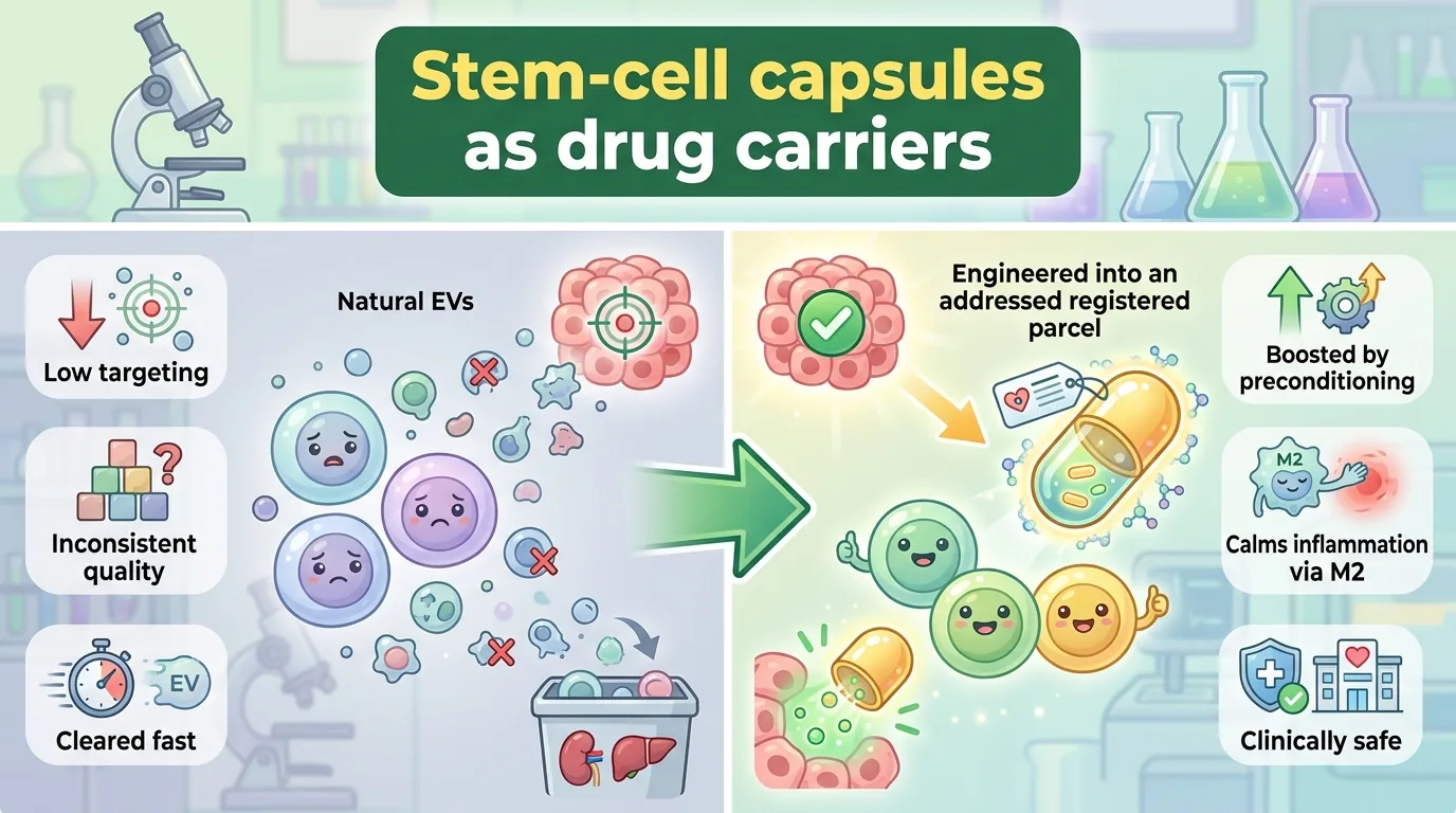
However, natural EVs had major weaknesses. First, natural MSC-EVs are weak at homing in on specific tissues; once administered into the body, they tend to rely on a passive distribution that “spreads and pools more or less at random,” so the efficiency with which they reach their target is limited. Second, the amount and variety of “cargo (drug)” that natural EVs can load hit a ceiling set by the state of the source stem cells. Third, it is difficult to reliably secure — using natural EVs — the quantity and purity needed for use in patients. In other words, “the material is good, but as-is it cannot clear the hurdle to practical use” — this has been the long-standing challenge.
Another fundamental question is “is it truly safe in the human body?” Even as its promise was being talked about, the painstaking safety-verification data — which doses and administration routes are safe in actual patients, whether side effects appear — were scattered, making the overall picture hard to grasp. This review attempts to answer these two questions — (1) how to remodel EVs into “usable carriers,” and (2) whether they were safe when tested in humans — by gathering studies from around the world.
What This Paper Reveals
What Is an EV — The Three Types of “Capsules” Cells Release
First, let’s lay out what EVs actually are. By how they form and their size, EVs are broadly divided into three types. Exosomes (40–160 nm) arise through an elaborate route: within the cell, a sac called a multivesicular body (MVB) is formed, and this fuses with the cell membrane and is released to the outside. Microvesicles (200–800 nm) form when the cell membrane buds directly outward and pinches off. And apoptotic bodies (1–5 μm) are the largest fragments, formed when a cell reaches the end of its life and dies by self-destruction (apoptosis). A nanometer (nm) is one-millionth of a millimeter. Exosomes belong to a world even smaller than one-thousandth the thickness of a human hair.
The EVs that MSCs put out carry an “owner’s name tag.” In addition to the tetraspanins common to many EVs (CD9, CD63, CD81), they bear the MSC markers CD44, CD73, CD90, and CD105, while lacking the blood-cell markers CD34 and CD45 — this combination lets you tell that “this is an MSC-derived EV.” As for their contents, they carry three kinds of information molecules — proteins, lipids, and nucleic acids (such as miRNAs) — and these rewrite the recipient cell’s behaviors such as proliferation, differentiation, and apoptosis (self-destruction).
The MSCs themselves, it turns out, can be obtained from a variety of “sources.” Adipose tissue, peripheral blood, bone marrow, dental pulp, placenta, umbilical cord, umbilical cord blood — the tissue of origin changes what the resulting EVs are good at. Bone marrow-derived MSCs (BM-MSCs) put out large amounts of neurotrophic factors such as BDNF and NGF, so they suit brain and spinal cord injuries; adipose-derived MSCs (AT-MSCs) suit skin (dermis and epidermis) regeneration and reproductive-system diseases; and umbilical cord-derived MSCs (UC-MSCs) have a strong immunomodulatory capacity and low immunogenicity, making them suited to lung diseases such as acute respiratory distress syndrome (ARDS). Choosing the “source” of the material is itself a fine first step in engineering.
Why Are MSC-EVs Well Suited to Being “Carriers”?
MSC-EVs draw attention as drug carriers (drug delivery carriers) because they combine the following advantages. Because they have a natural “shell” — the lipid bilayer — they blend easily into the body and are not readily beaten down by the immune system. They hold the latent power to cross the blood-brain barrier and reach the brain. Moreover, because the EVs themselves are loaded with proteins and miRNAs that act on inflammation and tissue repair, the capsule itself has a therapeutic effect, separate from the drug it carries. This point — that “the contents have drug efficacy from the start,” something synthetic nanoparticles lack — is a strength unique to MSC-EVs.
👦 Student: If they already have drug efficacy, why bother remodeling them at all?
🧬 Exotaro: Good question. They can work as-is, sometimes. But scattering large quantities of “letters with no address” is inefficient. To the targeted organ, in the targeted amount, reliably. That’s why we use “remodeling (engineering)” to upgrade a natural capsule into “registered mail with an address on it.”
Engineering: Six Ways to Turn a Plain Letter Into “Registered Mail With an Address”
The core of this review is an organization of the engineering strategies that boost the function of MSC-EVs. Broadly, they split into “preconditioning” — stimulating the cells in advance — and methods that work directly on the EVs, and they can be summarized into the following six categories.
👦 Student: The term “M2 type” keeps coming up from here on — what is it?
🧬 Exotaro: The macrophage — the “immune system’s cleanup crew” — has, broadly, two faces. The M1 type is the “attacking face” that fans the flames of inflammation, while the M2 type is the “healing face” that calms inflammation and repairs tissue. At the site of disease, inflammation tends to run out of control, so if you can switch cells from M1 to M2, putting out the fire and repair proceed at the same time. Much of the therapeutic effect of MSC-EVs can be explained by this “tilting toward M2” action. That’s why it will keep appearing from here on.
① Pharmacological preconditioning and drug loading. When you stimulate MSCs with a drug and then collect the EVs, the contents of the EVs change. For example, exosomes from MSCs preconditioned with Tanshinone IIA (TSA) ease myocardial ischemia-reperfusion injury via miR-223-5p. Preconditioning with melatonin increases IL-10 and Arg-1 via the PTEN/AKT pathway, tilting macrophages toward the inflammation-calming M2 type and promoting wound healing in diabetic mice. Bone marrow-derived MSC-EVs preconditioned with atorvastatin (ATV) advance M2 polarization via the miR-139-3p/Stat1 pathway, enhancing cardiac repair in acute myocardial infarction. Preconditioning with dexamethasone (Dex) suppresses genes involved in the inflammatory M1 type and increases the anti-inflammatory M2 type. Furthermore, EVs can also have “a drug loaded into them afterward”: bone marrow MSC exosomes loaded with the anticancer drug doxorubicin (Exo-Dox) enhanced antitumor efficacy while curbing toxicity in osteosarcoma, and kartogenin (KGN) for osteoarthritis (OA), when used intra-articularly together with exosomes modified with the MSC-targeting E7 peptide, significantly improved treatment outcomes over KGN alone. In addition, MSC-EVs loaded with the anti-VEGF drug bevacizumab reduced the number of intravitreal injections needed in diabetic retinopathy, lightening the patient’s burden, and MSC exosomes have been reported to carry the active ingredient of a herbal medicine to the brain, promoting autophagy in an Alzheimer’s disease model mouse and improving cognitive and motor function. The key point of this category is that by combining preconditioning with drug loading, the anti-inflammatory, repair, and regenerative power of natural EVs can be boosted even further.
② Genetic modification. This is a method that introduces a gene to rewrite the “message” loaded onto the EV. Exosomes from MSCs into which GDNF (glial cell line-derived neurotrophic factor) was introduced enhanced repair of kidney injury, and MSC-EVs in which SP1 was edited with CRISPR/Cas9 showed a protective effect against renal ischemia-reperfusion injury. Activating the anti-inflammatory protein TSG-6 with CRISPR technology increases the power to suppress inflammation, and conversely you can use it to “erase” a targeted gene. Bone marrow MSC-EVs loaded with radioiodine-labeled Cas9 (¹²⁴I@EVs-Cas9) suppressed the potassium channel KCNJ2 in osteosarcoma to halt proliferation and metastasis, and exosomes modified with a cartilage-adhering peptide (Cap) delivered CRISPR/Cas9 to the chondrocytes of OA patients to precisely knock out the ASPN gene. What is interesting is that MSC-EVs can also serve as excellent gene-editing carriers, protecting the CRISPR/Cas9 “components themselves” from degradation as they transport them. A natural capsule protects a fragile editing tool while sending it into the cell — a use unique to EVs.
③ Hypoxia treatment. When you grow MSCs with the oxygen deliberately thinned (a representative condition being 1–5%, considered physiological hypoxia, for 24–48 hours), the cells flip on a self-defense switch and put out EVs rich in factors that promote anti-inflammation, anti-apoptosis, and angiogenesis. The key player is HIF-1α, induced by hypoxia. What this review highlights especially in the neural field is the finding that EVs from hypoxia-preconditioned MSCs (HS-EVs) carried miR-146a-5p to steer macrophages toward the M2 type and suppress oxidative stress, easing inflammation and injury after spinal cord injury (SCI). In another study, exosomes from hypoxia-preconditioned MSCs were reported to boost the recovery of motor function after SCI via the miR-21/JAK2/STAT3 pathway.
④ Cytokine stimulation (priming). When you stimulate MSCs in advance with inflammatory cytokines such as TNF-α and IFN-γ, you obtain EVs with a strong immunomodulatory capacity. Giving TNF-α and IFN-γ together increases the secretion-related molecule RAB27B, so that large amounts of immunomodulatory EVs are produced. Also, exosomes from MSCs preconditioned with another cytokine, interleukin-1β (IL-1β), were reported to load abundant miR-21 and, in a sepsis model, promote M2 polarization of macrophages to improve symptoms. What is noteworthy is that this preconditioning reduces “donor-to-donor variability.” Single-cell analysis showed that even MSCs whose properties differ from person to person “converge” on a similar immunomodulatory program under TNF-α/IFN-γ stimulation, and it is regarded as a promising strategy toward manufacturing products of consistent specification. However, because it is an inflammatory stimulus, there is a possibility of carrying undesirable molecules, and this review cautions that careful optimization of conditions is needed.
⑤ Hybrid EVs. Fusing the “blend-in-ability” of natural EVs with the “ease of design” of artificial nanoparticles yields the hybrid membrane nanovesicle (HMNV). Mixing EVs with lipid nanoparticles (LNPs) lowered cytotoxicity compared with LNPs alone, and combining EVs with PLGA nanoparticles produced a sustained-release system that lets hUC-MSC exosomes act at the affected site for a long time. MSC-Hyb nanoparticles made with microfluidics and ultrasound delivered type I collagen mRNA — too large for natural EVs to load fully — to tendon stem cells, breaking through the loading limit of EVs. Also, hybrid vesicles modified with a dual-targeting peptide aimed at bone and muscle delivered miR-206-5p to suppress DUSP4 and activate the p38 MAPK pathway, promoting bone and muscle differentiation and showing efficacy in a mouse muscle-atrophy model. This direction takes the “best of both” natural and artificial to make up for weaknesses such as delivery stability and loading capacity.
⑥ Three-dimensional culture and physical stimulation. When you grow MSCs in three dimensions rather than on a flat (two-dimensional) surface, EV secretion increases roughly twofold, and the contents also change to become rich in anti-inflammatory and M2-inducing components. Physical stimulation works too. Low-intensity pulsed ultrasound (LIPUS) raised secretion of bone marrow MSC-EVs 3.66-fold, and electrical stimulation and light stimulation (photobiomodulation) have also been shown to boost the quantity and function of EVs. In short, you give the cells “moderate stress” to make them put out large amounts of good-quality EVs.
When Actually Tested in Humans, Was It Safe?
However glamorous the talk of remodeling, in the end it all comes down to “is it safe to use in humans?” The latter half of this review carefully takes stock of the clinical data (safety) for MSC-EVs across diseases.
In lung diseases, COVID-19-related acute respiratory distress syndrome (ARDS) has been the most studied. In a Phase II trial (102 people) of the bone marrow MSC-EV product “ExoFlo,” intravenous administration was tested, and in a 2024 double-blind randomized trial (21 people) that administered human placenta-derived MSC small EVs (hPMSC-sEV) at 1.5–2×10⁹ particles per kg of body weight for two days, oxygenation indices improved with no serious adverse events. In chronic obstructive pulmonary disease (COPD), inhalation of placenta-derived MSC exosomes eased inflammation and emphysema and improved patients’ quality of life (QOL). In idiopathic pulmonary fibrosis, hUC-MSC-EVs (24 people) administered by nebulizer (inhalation) were reported to significantly improve forced vital capacity (FVC) and 6-minute walk distance. Especially important is the result that in a meta-analysis of individual patient data pooling controlled trials, MSC-EV administration significantly lowered the odds ratio of death to 0.46 (95% confidence interval 0.26–0.81).
In skin diseases, examples cited include skin rejuvenation using adipose-derived MSC (AT-MSC) exosomes in combination with microneedling; the fact that using Wharton’s jelly-derived MSC-EVs in patients with atopic dermatitis produced no harmful local or systemic reactions (a confirmation of safety); the fact that AT-MSC exosomes eased refractory facial eczema that had arisen from the atopy drug dupilumab; and the fact that in a 110-person trial in diabetic foot ulcer (DFU), local administration of exosomes once a week for four weeks accelerated wound healing (with no serious adverse events).
Neurological diseases are squarely at the center of my (Exotaro’s) interest. In mild-to-moderate Alzheimer’s disease, a Phase I/II trial (9 people) of intranasal administration of AT-MSC exosomes was safe, and at the medium dose an improvement in the cognitive function score (ADAS-cog) was seen. In traumatic brain injury, a Phase I (5 people, 30 billion exosomes) improved motor function, cognition, and quality of life, and in spinal cord injury, a Phase I trial (9 people) of intrathecal administration of hUC-MSC exosomes was safe and was shown to potentially be associated with functional improvement.
Furthermore, clinical application is being attempted across a truly broad range of areas: the eye (refractory macular hole, dry eye due to GVHD), the gut (refractory perianal fistula in Crohn’s disease), the kidney (in stage III/IV chronic kidney disease, administration of hUC-MSC-EVs at 100 μg/kg improved kidney function and inflammation), and bone and joints (in osteoarthritis, intra-articular injection of hUC-MSC exosomes with no adverse events; MSC-derived apoptotic bodies (MSC-apoV) effective for hemostasis and bone regeneration).
Not to be overlooked in discussing safety are the dose-escalation trials conducted in healthy people. Nebulized inhalation of adipose-derived MSC-EVs was given as a single dose to 24 healthy volunteers, and tolerability was good even when divided into five levels from a low to a high group (from 2.0×10⁸ to 16×10⁸ particles). In a 10-person trial in which MSC exosomes were applied to the forearm skin three times a day for 20 days, no serious adverse events were reported either. Painstakingly verifying how far the safety margin extends not only in sick people but in healthy people — it lacks flash, but it is the indispensable accumulation for practical use. On the whole, the current data support the clinical safety of MSC-EVs and their dose-dependent safety (within a certain range, increasing the dose is fine). However, the review candidly acknowledges limitations such as the still-small number of cases, individual variation, and concurrent use with other drugs.
How Does It Differ From Synthetic Nanoformulations (Liposomes and the Like)?
This review compares MSC-EVs head-on with liposomes, polymeric nanoparticles, and lipid nanoparticles (LNPs). The strengths of MSC-EVs are their high biocompatibility, low immunogenicity, and ability to cross the blood-brain barrier — all owing to their natural origin — plus the “drug efficacy of the contents themselves” in the form of proteins and miRNAs. Liposomes and LNPs, on the other hand, lead with decades of clinical track record and with ease of achieving batch-to-batch quality consistency and mass production. Put the other way around, MSC-EVs are a case of “the material is the strongest, but quality uniformity, mass production, and standardization are the homework ahead.” That is precisely why the hybrid strategy of fusing natural EVs with artificial nanoparticles (⑤ above) is positioned as a promising direction that combines the best of both.
How Will the Future Change? (The Path to the Clinic)
Alongside the bright material, this review also confronts us with the “sober reality” of practical use. In one 2025 systematic review, 25 clinical trials using cell-derived EVs were identified; of these, the most numerous were COVID-19/ARDS (8 trials, 32%), followed by wound healing (5 trials, 20%), while trials with a control group numbered only 7 (28%). The neural field in particular is centered on Phase I, and large-scale Phase II/III trials for stroke and spinal cord injury remain a “gaping blank.”
👦 Student: If it’s been shown to be safe, can’t it be used as a treatment already?
🧬 Exotaro: I understand the feeling. But “safe” and “effective” are separate matters. Phase I is mainly the stage for confirming safety; to prove that something truly works, you have to pass controlled trials that compare against a placebo — Phase II/III. Right now, controlled trials still make up less than 30% of the total. The foundation of safety has been solidly built up, so next comes, at last, the phase of “rigorously demonstrating efficacy.” This is the crucial juncture.
There are mainly three remaining challenges. First, heterogeneity. EVs vary in how they form, their size, and their contents, and on top of that the source MSCs shift in character with each donor, tissue, and culture passage. Second, standardization. There is no unified specification for preparation and purification, so quantity, quality, and function vary from trial to trial, and moreover contamination by impurities could impair drug efficacy. Third, storage, transport, and cost. Keeping EVs stable requires strict low-temperature, sterile management, which drives up the cost of mass manufacturing. In addition, distribution and metabolism (pharmacokinetics) in the body have not yet been sufficiently elucidated. Intravenously injected EVs are eaten by resident macrophages and vanish quickly from the blood, pooling mainly in the liver and spleen. According to one systematic review, after intravenous injection there is first a distribution peak in the liver and kidney in the first hour, and over the following 2–12 hours it shifts to the lung and spleen. Moreover, this distribution varies greatly with the administration route, cell source, culture conditions, and purification method. That is exactly why the optimal dose, frequency, and route have not yet been settled. Against this “vanishes quickly” weakness, measures such as surface PEGylation, introducing CD47 to evade phagocytosis, and wrapping EVs in a hydrogel to make them act at the affected site for a long time are advancing in preclinical work. The administration route itself also governs how it works, and the idea of choosing “an entry point matched to the organ you want to reach” becomes important — intranasal to the brain, inhalation to the lung, local injection to keep it at the affected site.
The regulatory framework is also coming together. The European Union evaluates EV medicines within the framework of the advanced therapy medicinal product (ATMP, EU Regulation 1394/2007); the US FDA treats them as biologics and emphasizes CMC data (chemistry, manufacturing, and controls) and IND applications; and China’s NMPA is also incorporating them into the ATMP framework to advance full-lifecycle management. Internationally, a worldwide harmonization of standards through the ICH (International Council for Harmonisation) is anticipated. This review lists as homework for the future: optimizing the administration route (inhalation, microneedle, local injection, and so on) according to the characteristics of the disease and the pharmacokinetics; solidifying the evidence with large-scale, multicenter, randomized controlled trials; and establishing a quantitative quality specification (such as a specific protein profile) that can definitively state “this is an MSC-EV.”
Exotaro’s Perspective
I myself have researched how to treat neurological diseases, spinal cord injury foremost among them, using mesenchymal stem cells and extracellular vesicles. So this review is not one I can regard as someone else’s concern.
What resonated most with me is the view that “the quality of the material” and “the design of delivery” are separate things. Natural MSC-EVs are a rare material that itself possesses “the drug efficacy of the contents” — anti-inflammation and miRNAs. Yet there is a world of difference in the quality of the treatment that reaches the patient between scattering them as-is and leaving them to vanish into the liver and spleen, versus preconditioning them with hypoxia or cytokines to optimize the contents and designing the “delivery” itself through hybridization or hydrogels. It resonated straight through with the theme I usually care about — “how to deliver” rather than “how much to load.”
And the observation that the neural field is centered on Phase I, with large-scale controlled trials for spinal cord injury and stroke left blank, is at once a hope and a piece of homework. The step that this review cites — that the Phase I of intrathecal hUC-MSC exosomes in spinal cord injury “was safe” — is small but certain, and I feel it is my responsibility, as one person working in this field, to carry it forward into trials that rigorously demonstrate the efficacy that lies beyond. To the natural capsule, we add just a little wisdom. Believing that this accumulation will one day reach a future where “a person who could not walk stands up again,” I continue my research today as well.