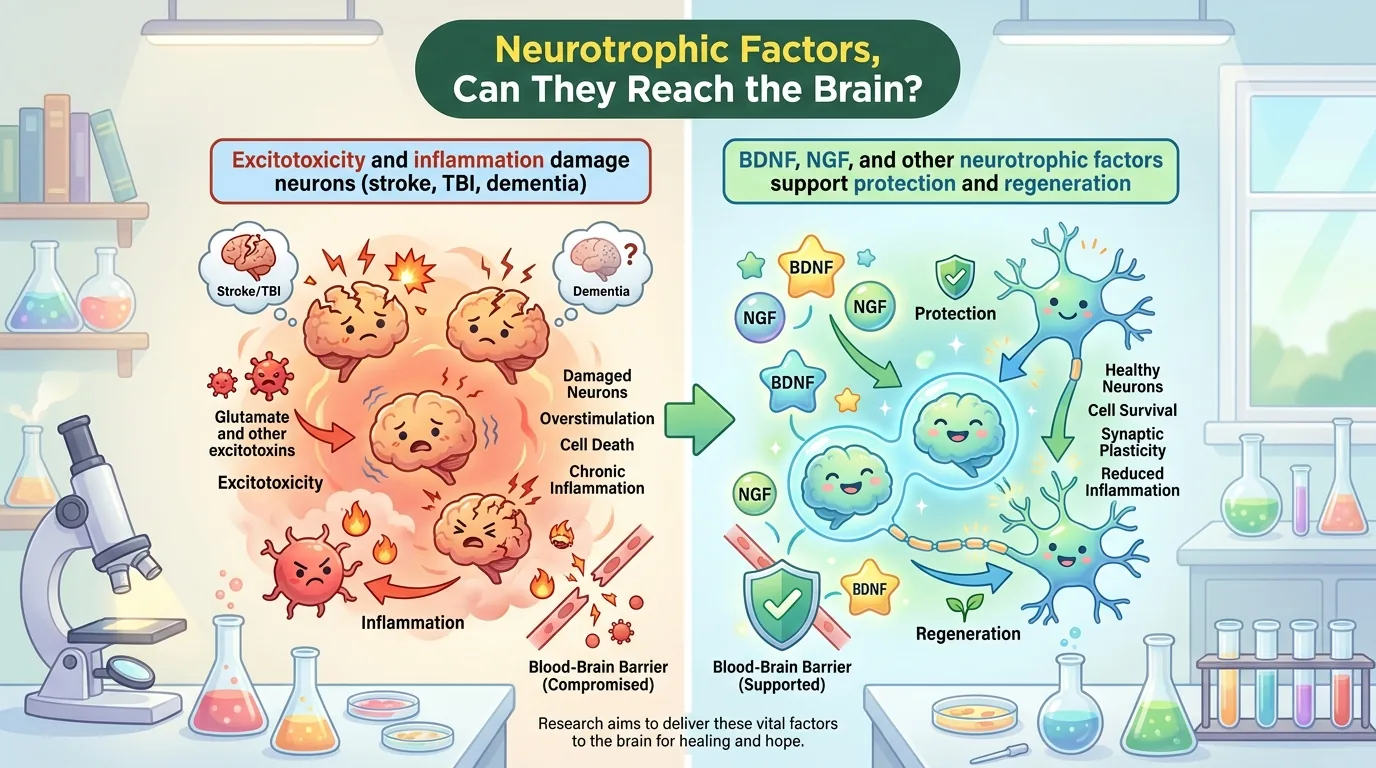
The brain in the moments right after a stroke, the brain in the moments right after a traumatic impact, and the brain of Alzheimer’s disease or Parkinson’s disease, where neurons die slowly over decades—the triggers are completely different, yet at the cellular level, we are finding that these brains break down in remarkably similar ways. Excitotoxicity, oxidative stress, mitochondrial dysfunction, neuroinflammation, and the breakdown of the blood-brain barrier (BBB), the checkpoint that protects the brain. The molecules that have long drawn attention as the protagonists standing up to this shared “mode of breakdown” are neurotrophic factors (NTFs). A review paper compiled by a research team at Belgorod State National Research University in Russia surveys everything from the biology of NTFs to actual clinical trial data, attempting to answer the core question: why do drugs that should work fail to help patients?
Publication Details
- Paper title: Neurotrophic Factors in Stroke, Traumatic Brain Injury, and Neurodegeneration: A Convergent Pathophysiological and Translational Perspective
- Paper type: Review article. By the authors’ own description, this is a narrative review that synthesizes original research articles, systematic reviews, and meta-analyses published over roughly the past 10 years, along with classic literature that laid the foundation for neurotrophic factor research. It is not a systematic review.
- Authors: Olesya V. Shcheblykina (corresponding author), Darya A. Kostina, Mikhail V. Pokrovskii, Mikhail V. Korokin
- Affiliation: Research Institute of Pharmacology of Living Systems, Belgorod State National Research University, Russia
- Journal: Journal of Integrative Neuroscience (IMR Press), 2026, Volume 25, Issue 7, Article No. 51543
- DOI / Link: 10.31083/JIN51543
- Peer review and publication dates: Submitted March 5, 2026; revised April 29; accepted May 8; published July 15. Handling editors were Maarten Van den Buuse and Bettina Platt.
- Open access: Yes (CC BY 4.0. Free to reuse and redistribute with attribution)
- Impact Factor: 2.7 (Clarivate’s 2024 Journal Citation Reports, published June 18, 2025; ranked 174th out of 314 journals in Neuroscience, Q3). The figure rose further in the latest JCR release (2025 data), published June 2026, with the publisher’s website now listing “3.3 (2025)” and “Q2” as the current values. The journal is indexed in SCIE (Web of Science), MEDLINE (PubMed), Scopus, and DOAJ.
- About the journal: According to the publisher’s own description, it is an open-access journal covering every domain of neuroscience, from the molecular, cellular, and systems levels through to translational research. Its Scopus-based CiteScore is 5.
- Funding: Ministry of Science and Higher Education of the Russian Federation, State Assignment No. FZWG-2026-0003
- Conflicts of interest: The authors declare no conflicts of interest.
About corresponding author Olesya V. Shcheblykina: The corresponding author is a researcher at the Research Institute of Pharmacology of Living Systems at Belgorod State National Research University, and also holds a teaching post as Associate Professor (доцент, Associate Professor) in the university’s Department of Pharmacology and Clinical Pharmacology within the Faculty of Medicine. They belong to the same institute as co-authors Pokrovskii and Korokin, and the three have repeatedly collaborated on multiple neuroscience papers from that institute beyond this one. For example, in 2025 they published a study on sex differences and histomorphological biomarkers using an Alzheimer’s disease mouse model (APPswe/PS1dE9/Blg strain) (in Brain Sciences), and in 2026 a study examining how traumatic brain injury affects synuclein-related transcription, amyloid plaque morphology, and cognitive function (in Biomedicines), suggesting that experimental neuropharmacology spanning neurodegenerative disease and brain injury is their area of specialization. Given the consistency of these research themes, this review appears to be built on the corresponding author’s own experimental research interests. Note that the corresponding author’s ORCID (0000-0003-0346-9835) still lists an outdated affiliation of “graduate student” as of September 2017, but Belgorod State National Research University’s official faculty roster already lists the title of Associate Professor, which is likely the more current information.
What Wasn’t Understood Before?
The adult central nervous system (CNS) is a tissue with an extremely limited capacity to rebuild neural circuits on its own once they are damaged. That is precisely why stroke, traumatic brain injury (TBI), and neurodegenerative diseases (NDDs)—represented by Alzheimer’s disease (AD) and Parkinson’s disease (PD)—are all so difficult to recover from once neurons are lost. What this review demonstrates first is the fact that these diseases, despite having entirely different causes and speeds of onset, follow remarkably similar pathways to destroy neurons at the cellular and molecular level.
In ischemic stroke, for example, when blood flow stops and cells can no longer produce ATP, the cell’s energy currency, neurons lose the ability to maintain their membrane potential, and the excitatory neurotransmitter glutamate leaks out in large quantities. This over-stimulates NMDA receptors, triggering a flood of calcium ions into the cell—a phenomenon known as “excitotoxicity.” Excess calcium sends degrading enzymes such as calpain and phospholipase A2 into overdrive, damaging the cytoskeleton, cell membrane, and DNA. At the same time, nitric oxide synthase (NOS) becomes overactive, and nitric oxide (NO) combines with superoxide (O2⁻) to form the highly toxic peroxynitrite (ONOO⁻), which strikes the mitochondria directly and further increases reactive oxygen species (ROS). Microglia gather around the damaged neurons and release inflammatory cytokines such as TNF-α, IL-1β, and IL-6, spreading the damage further—ultimately, neurons are lost to necrosis at the core of the infarct, and to apoptosis (programmed cell death) in the surrounding region known as the “penumbra.”
In hemorrhagic stroke, blood itself (breakdown products of hemoglobin) is directly toxic, and secondary ischemia from elevated intracranial pressure also comes into play, so the inflammatory response is generally more severe than in ischemic stroke. In TBI, the primary injury from the impact itself is followed by secondary injury—a cascade of blood-brain barrier breakdown, osmotic disturbance, inflammation, and oxidative stress. And in neurodegenerative diseases such as Alzheimer’s disease, Parkinson’s disease, and amyotrophic lateral sclerosis (ALS), the trigger is not an acute accident but the slow accumulation of abnormal proteins—amyloid-β and tau protein (AD), or α-synuclein (PD)—yet the cellular response that follows—excitotoxicity, oxidative stress, neuroinflammation, apoptosis—is, the authors note, “strikingly similar” to that seen in stroke and TBI.
👦 Student: The causes are all different, but the way things break down is almost the same?
🧬 Dr. Exotaro: That’s right. It’s a bit like how, whether a fire starts from an electrical short or arson, the way it spreads and the basic principles for putting it out are the same. That’s exactly why drugs that stop the “shared mode of breakdown”—rather than the cause itself—neurotrophic factors, have drawn attention for years as treatment targets that cut across multiple diseases.
It is also known that, in a brain undergoing this kind of breakdown, the expression and availability of the endogenous neurotrophic factors that support neuronal survival, plasticity, and regeneration drop substantially. If neurotrophic factors could be supplemented from outside, or their synthesis promoted, they would help produce anti-apoptotic proteins, improve synaptic plasticity, promote neurogenesis, switch microglia from an inflammatory to a neuroprotective phenotype, and act as antioxidants as well—in theory, they have long been considered a potential “universal treatment target” spanning stroke, TBI, and neurodegenerative disease.
What Did This Paper Find?
Research into neurotrophic factors traces back to the 1950s and the discovery of NGF. Rita Levi-Montalcini and Viktor Hamburger identified nerve growth factor (NGF) from the observation that transplanting mouse tumors into chick embryos caused nerve fibers to sprout vigorously, and Levi-Montalcini received the 1986 Nobel Prize in Physiology or Medicine. Brain-derived neurotrophic factor (BDNF) was subsequently isolated, and gene cloning revealed it was structurally similar to NGF, giving rise to the concept of a “neurotrophic factor family.” Neurotrophin-3 (NT-3) and neurotrophin-4 (NT-4) were later added to the family, and neurotrophin-6 and -7 have also been found in fish, though these have no mammalian homologs.
Mature NTFs are dimers formed from a pair of molecules, stabilized by a structure called a “cystine knot” made of three disulfide bonds. NTFs are a broad family that includes not only narrowly-defined neurotrophins such as NGF, BDNF, and NT-3, but also GDNF, CNTF, insulin-like growth factor (IGF), basic fibroblast growth factor (bFGF), vascular endothelial growth factor (VEGF), TGF-β, and Sonic hedgehog (Shh). There are mainly two receptor systems—the high-affinity tropomyosin receptor kinase (Trk) family, and the low-affinity p75 neurotrophin receptor (p75NTR), which belongs to the TNF receptor superfamily. NGF binds TrkA, BDNF and NT-4 bind TrkB, and NT-3 mainly binds TrkC, activating the Ras-MAPK and PI3K-Akt pathways. p75NTR, meanwhile, can bind every neurotrophic factor, but has a dual nature: in the absence of Trk receptors, it can instead act to induce apoptosis.
One point this review discusses with particular emphasis is that NTF function changes substantially with age and sex. In a study using human post-mortem brain tissue (orbitofrontal cortex, n=209, ages 16-96), BDNF mRNA levels declined progressively with age, in tandem with reduced expression of 78 genes involved in excitatory and inhibitory synaptic function. Even more severe changes have been reported for NGF. In the prefrontal cortex and hippocampus of aged rats (19-22 months old), the immature form, proNGF, increased 1.8- to 1.9-fold, while mature NGF decreased by as much as 36-44%. At the same time, p75NTR increased 1.6- to 1.8-fold and the co-receptor sortilin increased 1.8- to 2.1-fold, causing p75NTR—normally neuroprotective—to “transform” into a high-affinity, apoptosis-inducing receptor for the accumulated proNGF.
👦 Student: So as you age, the trophic factor itself can become “toxic”?
🧬 Dr. Exotaro: Exactly. This doesn’t happen in young animals. It’s a bit like how a nutritional supplement that works wonders on a growing body can instead cause indigestion in a body with a slower metabolism. A treatment that simply adds trophic factors from outside might instead send a danger signal in elderly patients—that’s exactly why this review sounds a warning about the design of clinical trials centered on older patients.
With NT-3, what is even more interesting is that whether it works depends strictly not just on “age” but on “time elapsed since injury.” In a rat spinal cord injury model, adenoviral vector-mediated NT-3 overexpression strongly promoted corticospinal tract axon growth when administered in the acute phase, two weeks after injury, but had no effect when administered in the chronic phase, four months after injury. This is thought to be because a co-induction signal derived from Wallerian degeneration, present only in the acute phase, is lost by the chronic phase. It should be noted, however, that this finding is based on a comparison in a specific vector and injury model, and is not an absolute rule that can be generalized to every neurotrophic factor and route of administration.
Clinical data also support NTFs’ actual involvement. In patients with ischemic stroke, serum NGF concentration was in fact significantly higher than in healthy controls, and a clear inverse correlation was observed between NGF concentration and NIHSS (a score indicating stroke severity). Some reports also found that patients with higher serum NGF concentrations in the acute phase (at admission) had better functional outcomes (assessed by the modified Rankin Scale) at 3 months after onset. Significant decreases in BDNF have likewise been confirmed in patients with stroke and neurodegenerative disease, while VEGF is known to spike sharply in the blood after stroke onset—whether ischemic or hemorrhagic—and to remain elevated for at least 90 days. All of these findings hint at potential future use as biomarkers.
Building on this basic biology, the authors compare in detail the actual means of delivering these factors to the brain. Viral vectors (AAV), lipid nanoparticle-based mRNA delivery, extracellular vesicles (EVs) including exosomes, sustained release via hydrogels, cell therapy, small-molecule mimetics, and intranasal administration—the authors summarize 13 preclinical studies from 2016 to 2026 in a comparison table, concluding that AAV gene therapy, lipid nanoparticles, exosome-based systems, and transplantation of cells that continuously secrete NTFs produce the most convincing results, while small-molecule mimetics, though able to cross the blood-brain barrier, still lag behind in selectivity and magnitude of effect. Among these, intranasal delivery of mesenchymal stem cell (MSC)-derived extracellular vesicles (EVs) loaded with a BDNF-enhancing neuropeptide is positioned as a promising candidate that combines non-invasiveness with low immunogenicity. Delivering BDNF via neural stem cell-derived exosomes to a cerebral infarction model is also a promising approach with the advantage of low immunogenicity, though its invasive route of administration—stereotactic injection—is cited as a limitation.
And the core of this review is its summary of six clinical trials actually conducted in humans (Table 3): direct injection of AAV2-BDNF into the hippocampus and entorhinal cortex for Alzheimer’s disease (NCT05040217, Phase I, ongoing); bilateral intraputaminal administration of AAV2-GDNF for Parkinson’s disease (NCT06285643, Phase Ib-II); intermittent infusion of recombinant GDNF protein via an implanted pump (10 doses total at 4-week intervals, Phase II); intraputaminal administration of AAV2-neurturin (CERE-120) (NCT00985517, Phase I → Phase II/IIb); ex vivo NGF gene therapy using fibroblasts (Phase I, n=8); and direct injection of AAV2-NGF (NCT00087789 and NCT00017940, Phase I → Phase II)—while safety was generally reported as favorable in many of these trials, statistically significant effects on the primary endpoint were not demonstrated in most of them.
At the same time, positive findings have been reported at the level of target engagement and imaging or biological markers. In the ex vivo NGF gene therapy trial—a Phase I trial with a small sample (n=8) and no control group—cortical glucose metabolism was reported to increase significantly on PET imaging, and autopsy findings reported dense outgrowth of cholinergic axons into the graft. For AAV2-NGF, signs of sustained NGF expression and cholinergic activation were reported; for AAV2-GDNF, GDNF expression in dopaminergic neurons and stabilization of motor scores; and for AAV2-BDNF, preliminary data showing increased entorhinal cortex metabolism. The authors describe this as “a gap between molecular-level efficacy and clinical benefit,” and it is precisely this gap that underpins this review’s conclusion—that the problem lies not in the biology, but in the delivery platform.
The Phase II/IIb trial of CERE-120 in particular is singled out for “safety concerns,” and it also failed to show a significant difference from the sham-surgery group on the primary motor endpoint (UPDRS) at 12 months. In the paper’s limitations section, this trial is named specifically, with pointed language about “a high incidence of adverse events” and “a probable association with tumor formation.” In the intermittent infusion trial of recombinant GDNF as well, adverse events such as dyskinesia, paresthesia, Lhermitte’s sign, on-off phenomena, and diplopia occurred more frequently than in the placebo group (a difference of 3 or more cases between treatment arms). The authors condense this result into a single sentence in the paper: “no market-approved neurotrophic factor-derived pharmaceutical is currently used clinically for the treatment of neurodegenerative disease, stroke sequelae, or brain trauma.”
How Will the Future Change? (The Path to the Clinic)
The authors conclude that the main reason neurotrophic factor therapy has failed to clear the “wall” to practical use is not a lack of biological validity, but the absence of a delivery platform capable of getting these factors to the brain safely, effectively, and in a scalable form. Turn that around, and it means that every molecule this review has organized has ample grounds to serve as a treatment target—provided the delivery problem can be solved.
As concrete paths forward, this review points to the development of next-generation AAV capsids with enhanced CNS tropism, engineered exosome vectors, lipid nanoparticle platforms for delivering mRNA, and focused ultrasound technology that non-invasively and temporarily opens the blood-brain barrier. All of these aim at less invasive routes of administration to replace the current invasive practice of direct injection into the brain. In addition, the authors emphasize the necessity of developing inducible expression systems that can control expression levels, as well as patient stratification—narrowing down “who it will work for” based on disease stage, blood biomarker concentrations, and receptor genotype. In cases where delivering the full-length protein is difficult, small-molecule Trk receptor agonists and NTF mimetics are also positioned as pharmacologically tractable complementary strategies. Indeed, as the strict dependence of NT-3’s therapeutic window on time since injury shows, the effect of the same molecule can be diametrically opposite depending on “when” it is delivered.
👦 Student: Is refining “how it’s delivered” more important than improving the drug itself?
🧬 Dr. Exotaro: At least, that’s close to what this review concludes. We already know the contents of the trophic-factor “letter” are powerful enough. What’s missing is the postal system that delivers it to the right address, at the right time, in the right amount.
The authors also touch on the importance of an integrated approach that combines neurotrophic factor therapy with rehabilitation and standard drug therapy. The idea is that combining these factors, which protect and regenerate neurons on their own, with training that draws out plasticity, could produce synergistic functional recovery. As a conclusion, the future the authors envision is a shift from therapy that “simply replenishes a deficient trophic factor” to therapy that “precisely tunes neurotrophic factor signaling in space and time, matched to disease stage, individual biological background, and the characteristics of the delivery route.”
How to Critically Read This Research (Limitations and Paths to Improve It)
First, in fairness, it should be emphasized that the authors themselves disclose the limitations of this review in fairly concrete terms. The conclusion states explicitly that “most preclinical data were obtained from young adult male rodents under highly controlled experimental conditions, and do not adequately capture the biological diversity present in clinical populations.” Age-related decline in Trk receptor signaling, accumulation of pro-apoptotic molecules such as proNGF and proBDNF, impaired retrograde axonal transport, and persistent neuroinflammation—none of these are reproduced in standard animal models, and the authors themselves acknowledge that regulation of NTF expression and receptor sensitivity by sex hormones is also not adequately incorporated into experimental design. Given that the actual patient population for stroke and neurodegenerative disease is characterized by advanced age, vascular comorbidities, and polypharmacy, this gap cannot be dismissed lightly. From this data, the authors also state clear clinical implications. Summarized roughly, these are: (1) preclinical efficacy data obtained mainly from young adult male rodents may overestimate the neuroprotective effect actually obtained in elderly patients; (2) the age-related shift in signaling toward proBDNF and proNGF raises concern that exogenous NTF administration could instead activate the p75NTR-sortilin-dependent apoptotic pathway; (3) menopausal status and androgen levels should be considered as stratification variables in clinical trials targeting BDNF-dependent or dopaminergic circuits. These are not mere confessions of limitation but can be credited as concrete recommendations for the design of future trials.
👦 Student: What’s the concrete problem with “not being a systematic review”?
🧬 Dr. Exotaro: It’s a bit like a librarian saying, “I gathered 10 years’ worth of relevant books, in whatever order I happened to notice them.” The books they gathered might be high quality. But because there’s no record of which shelves they checked or which books they left out, there’s no way for another librarian to redo the same work later and confirm whether they’d arrive at the same conclusion. That’s the “reproducibility” problem.
On the other hand, the methodology of this review itself also needs to be addressed. This is not a systematic review but a narrative review. The selection policy stated explicitly in the text is limited to the description “original research articles, systematic reviews, and meta-analyses published over roughly the past 10 years, along with classic literature that laid the foundation”—no pre-registered review protocol, specific database names used for the search, inclusion/exclusion criteria, PRISMA flowchart, or meta-analytic method for pooling effect sizes are presented. This does not negate the review’s value as a review—it can in fact be a strength when it comes to providing a perspective that bridges a wide range of fields—but the limitation on reproducibility, in the sense that readers have no way to verify “which papers were selected and which were not,” should be honestly recognized. Possible directions for improvement include, for example, conducting a limited meta-analysis pooling effect sizes by delivery platform using only the Phase I-II human clinical trial data organized in Table 3, or incorporating a PRISMA-compliant protocol and GRADE evidence assessment into a future version to develop it into a systematic review.
It should also be noted that the authors themselves disclose that an AI-assisted tool (Perplexity) was used in drafting the manuscript, but its use is stated explicitly to have been limited to improving readability and proofreading grammar and notation, along with assisting with suggestions on the visual design of Figure 1, and not used to generate the content itself. The authors are stated to bear final responsibility for the accuracy and originality of the content, and this does not undermine the scientific credibility of the review.
Dr. Exotaro’s Perspective
Reading this review, the part that made me sit up was the NT-3 story. Delivering NT-3 via an adenoviral vector in the acute phase, two weeks after injury, produces corticospinal tract axon growth, yet doing the exact same thing in the chronic phase, four months later, has no effect—this is precisely the wall I run into every day in my own spinal cord injury (SCI) research using mesenchymal stem cell (MSC)-derived extracellular vesicles (EVs). The explanation that a co-induction signal tied to Wallerian degeneration exists only in the acute phase feels like it accurately captures part of the mechanism behind why the same intervention tends to lose its effect in chronic spinal cord injury models. That said, I also think it still needs to be carefully determined whether this is a universal law that extends beyond spinal cord injury or NT-3. Even so, I am struck once again by the lesson that “when it’s delivered” matters just as much as “what is delivered”—a principle common to both trophic factor research and EV research.
Further, the neuroprotection in a cerebral infarction model via BDNF-loaded neural stem cell-derived exosomes, the intranasal delivery method loading MSC-derived EVs with a neuropeptide, and stroke treatment using BDNF-producing MSCs embedded in a hydrogel—all covered in Table 2 of this review—are areas directly adjacent to my own research field. What these studies have in common is that they harness the low immunogenicity of exosomes and EVs, and their ability to cross the blood-brain barrier readily, as a means of transporting the “cargo” that is a trophic factor. Rather than simply injecting trophic factor protein, delivering it loaded onto the natural, nanoscale delivery system that cells already possess—I believe this idea directly addresses the problem this review repeatedly points to: that delivery is the greatest bottleneck.
As the clinical trial data show, an acceptable safety profile is being confirmed in many trials. That said, cases like the CERE-120 and intermittent GDNF infusion trials, where a high number of adverse events and a possible association with tumor formation were noted, still remain, and the safety story should not be oversimplified. What remains is the precision to deliver the right amount to the right patient, at the right time, safely. As someone working on spinal cord injury—a condition where “time since injury” quite literally determines the outcome—I want to carry forward the importance of this time window that this review raises, and weigh it against my own research going forward.